Antimetabolites


Antimetabolites


M. Wasif Saif

Edward Chu


ANTIFOLATES

Reduced folates play a key role in one-carbon metabolism, and they are essential for the biosynthesis of purines, thymidylate, and protein biosynthesis. Aminopterin was the first antimetabolite with documented clinical activity in the treatment of children with acute leukemia in the 1940s. This antifolate analog was subsequently replaced by methotrexate (MTX), the 4-amino, 10-methyl analog of folic acid, which remains the most widely used antifolate analog, with activity against a wide range of cancers (Table 19.1), including hematologic malignancies (acute lymphoblastic leukemia and non-Hodgkin’s lymphoma) and many solid tumors (breast cancer, head and neck cancer, osteogenic sarcoma, bladder cancer, and gestational trophoblastic cancer).

Pemetrexed is a pyrrolopyrimidine, multitargeted antifolate analog that targets multiple enzymes involved in folate metabolism, including thymidylate synthase (TS), dihydrofolate reductase (DHFR), glycinamide ribonucleotide (GAR) formyltransferase, and aminoimidazole carboxamide (AICAR) formyltransferase.1,2 This agent has broad-spectrum activity against solid tumors, including malignant mesothelioma and breast, pancreatic, head and neck, non-small-cell lung, colon, gastric, cervical, and bladder cancers.3,4,5

The third antifolate compound to have entered clinical practice is pralatrexate (10-propargyl-10-deazaaminopterin), a 10-deazaaminopterin antifolate that was rationally designed to bind with higher affinity to the reduced folate carrier (RFC)-1 transport protein, when compared with MTX, leading to enhanced membrane transport into tumor cells. It is also an improved substrate for the enzyme folylpolyglutamyl synthetase (FPGS), resulting in enhanced formation of cytotoxic polyglutamate metabolites.6,7 When compared with MTX, this analog is a more potent inhibitor of multiple enzymes involved in folate metabolism, including TS, DHFR, and GAR and AICAR formyltransferases. This agent is presently approved for the treatment of relapsed or refractory peripheral T-cell lymphomas.8


Mechanism of Action

The antifolate compounds are tight-binding inhibitors of DHFR, a key enzyme in folate metabolism.1 DHFR plays a pivotal role in maintaining the intracellular folate pools in their fully reduced form as tetrahydrofolates, and these compounds serve as one-carbon carriers required for the synthesis of thymidylate, purine nucleotides, and certain amino acids.

The cytotoxic effects of MTX, pemetrexed, and pralatrexate are mediated by their respective polyglutamate metabolites, with up to 5 to 7 glutamyl groups in a γ-peptide linkage. These polyglutamate metabolites exhibit prolonged intracellular half-lives, thereby allowing for prolonged drug action in tumor cells. Moreover, these polyglutamate metabolites are potent, direct inhibitors of several folate-dependent enzymes, including DHFR, TS, AICAR formyltransferase, and GAR formyltransferase.1


Mechanisms of Resistance

The development of cellular resistance to antifolates remains a major obstacle to its clinical efficacy.9,10 In experimental systems, resistance to antifolates arises from several mechanisms, including an alteration in antifolate transport because of either a defect in the reduced folate carrier or folate receptor systems, decreased capacity to polyglutamate the antifolate parent compound through either decreased expression of FPGS or increased expression of the catabolic enzyme γ-glutamyl hydrolase, and alterations in the target enzymes DHFR and/or TS through increased expression of wild-type protein or overexpression of a mutant protein with reduced binding affinity for the antifolate. Gene amplification is a common resistance mechanism observed in various experimental systems, including tumor samples from patients. In in vitro and in vivo experimental model systems, the levels of DHFR and/or TS protein acutely increase after exposure to MTX and other antifolate compounds. This acute induction of target protein in response to drug exposure is mediated, in part, by a translational regulatory mechanism, which may represent a clinically relevant mechanism for the acute development of cellular drug resistance.


Clinical Pharmacology

The oral bioavailability of MTX is saturable and erratic at doses greater than 25 mg/m2. MTX is completely absorbed from parenteral routes of administration, and peak serum levels are achieved within 30 to 60 minutes of administration.

The distribution of MTX into third-space fluid collections, such as pleural effusions and ascitic fluid, can substantially alter MTX pharmacokinetics. The slow release of accumulated MTX from these third spaces over time prolongs the terminal half-life of the drug, leading to potentially increased clinical toxicity. It is advisable to evacuate these fluid collections before treatment and monitor plasma drug concentrations closely.

Renal excretion is the main route of drug elimination, and this process is mediated by glomerular filtration and tubular secretion. About 80% to 90% of an administered dose is eliminated unchanged in the urine. Doses of MTX, therefore, should be reduced in proportion to reductions in creatinine clearance. Renal excretion of MTX is inhibited by probenecid, penicillins, cephalosporins, aspirin, and nonsteroidal anti-inflammatory drugs.

Pemetrexed enters the cell via the RFC system and, to a lesser extent, by the folate receptor protein. As with MTX, it undergoes polyglutamation within the cell to the pentaglutamate form, which is at least 60-fold more potent than the parent compound. This agent is mainly cleared by renal excretion, and in the setting of renal dysfunction, the terminal drug half-life is significantly prolonged to up to 20 hours. Pemetrexed, therefore, should be used with caution in patients with renal dysfunction. In addition, renal excretion is inhibited in the presence of other agents including probenecid, penicillins, cephalosporins, aspirin, and nonsteroidal anti-inflammatory drugs.


TABLE 19.1 Antimetabolites: Indications, Doses and Schedules, and Toxicities


Drug


Main Therapeutic Uses


Main Doses and Schedule


Major Toxicities


Methotrexate


Non-Hodgkin’s lymphoma


Primary CNS lymphoma


Acute lymphoblastic leukemia


Breast cancer


Bladder cancer


Osteogenic sarcoma


Gestational trophoblastic cancer


Low dose: 10-50 mg/m2 IV every 3-4 weeks


Low dose weekly: 25 mg/m2 IV weekly


Moderate dose: 100-500 m/m2 IV every 2-3


weeks High dose: 1-12 gm/m2 IV over a 3- to 24-hour period every 1-3 weeks


Intrathecal (IT): 10-15 mg IT 2 times weekly until CSF is clear, then weekly dose for 2-6 weeks, followed by monthly dose


Mucositis, diarrhea, myelosuppression, acute renal failure, transient elevations in serum transaminases and bilirubin, pneumonitis, neurologic toxicity


Pemetrexed


Mesothelioma


Non-small-cell lung cancer


500 mg/m2 IV, every 3 weeks


Myelosuppression, skin rash, mucositis, diarrhea, fatigue


Pralatrexate


Peripheral T-cell lymphoma


30 mg/m2 IV, weekly for 6 weeks; cycles repeated every 7 weeks


Myelosuppression, skin rash, mucositis, diarrhea, elevation of serum transaminases and bilirubin, mild nausea/vomiting


5-Fluorouracil


Breast cancer


Colorectal cancer


Anal cancer


Gastroesophageal cancer


Hepatocellular cancer


Pancreatic cancer


Head and neck cancer


Bolus monthly schedule: 425-450 mg/m2 IV on days 1-5 every 28 days


Bolus weekly schedule: 500-600 mg/m2 IV every week for 6 weeks every 8 weeks


Infusion schedule: 2,400-3,000 mg/m2 IV over 46 hours every 2 weeks


120-hour infusion: 1,000 mg/m2/d IV on days 1-5 every 21-28 d


Protracted continuous infusion: 200-400 mg/m2/d IV


Nausea/vomiting, diarrhea, mucositis, myelosuppression, neurotoxicity, coronary artery vasospasm, conjunctivitis


Capecitabine


Breast cancer


Colorectal cancer


Gastroesophageal cancer


Hepatocellular cancer


Pancreatic cancer


Recommended dose for monotherapy is 1,250 mg/m2 PO bid for 2 weeks with 1 wk rest


May decrease dose of capecitabine to 850-1,000 mg/m2 bid on days 1-14 to reduce risk of toxicity without compromising efficacy


An alternative dosing schedule for monotherapy is 1,250-1,500 mg/m2 PO bid for 1 week on and 1 week off; this schedule appears to be well tolerated, with no compromise in clinical efficacy


Capecitabine should be used at lower doses (850-1,000 mg/m2 bid on days 1-14) when used in combination with other cytotoxic agents, such as oxaliplatin and lapatinib


Diarrhea, hand-foot syndrome, myelosuppression, mucositis, nausea/vomiting, neurologic toxicity, coronary artery vasospasm


Cytarabine


Hodgkin’s lymphoma


Non-Hodgkin’s lymphoma


Acute myelogenous leukemia


Acute lymphoblastic leukemia


Standard dose: 100 mg/m2/day IV on days 1-7 as a continuous IV infusion, in combination with an anthracycline as induction chemotherapy for acute myelogenous leukemia


High-dose: 1.5-3.0 gm/m2 IV q 12 hours for 3 days as a high dose, intensification regimen for acute myelogenous leukemia


SC: 20 mg/m2 SC for 10 days per month for 6 months, associated with IFN-α for treatment of chronic myelogenous leukemia


IT: 10-30 mg IT up to 3 times weekly in the treatment of leptomeningeal carcinomatosis secondary to leukemia or lymphoma.


Nausea/vomiting, myelosuppression, cerebellar ataxia, lethargy, confusion, acute pancreatitis, drug infusion reaction, hand-foot syndrome


High-dose therapy: noncardiogenic pulmonary edema, acute respiratory distress and Streptococcus viridans pneumonia, conjunctivitis, and keratitis


Gemcitabine


Pancreatic cancer


Non-small-cell lung cancer


Breast cancer


Bladder cancer


Hodgkin’s lymphoma


Ovarian cancer


Soft tissue sarcoma


Pancreatic cancer: 1,000 mg/m2 IV every week for 7 weeks with 1 week rest Treatment then continues weekly for 3 weeks followed by 1 week off


Bladder cancer: 1,000 mg/m2 IV on days 1, 8, and 15 every 28 days


Non-small-cell lung cancer: 1,000-1,200 mg/m2 IV on days 1 and 8 every 21 days


Nausea/vomiting, myelosuppression, flulike syndrome, elevation of serum transaminases and bilirubin, pneumonitis, infusion reaction, mild proteinuria, and rarely, hemolytic-uremic syndrome and thrombotic thrombocytopenic purpura


6-Mercaptopurine


Acute lymphoblastic leukemia


Induction therapy: 2.5 mg/kg PO daily


Maintenance therapy: 1.5-2.5 mg/kg PO daily


Myelosuppression, nausea/vomiting, mucositis and diarrhea, hepatotoxicity, immunosuppression


6-Thioguanine


Acute myelogenous leukemia


Acute lymphoblastic leukemia


Induction: 100 mg/m2 PO every 12 hours on days 1-5, usually in combination with cytarabine


Maintenance: 100 mg/m2 PO every 12 hours on days 1-5, every 4 weeks, usually in combination with other agents


Single agent: 1-3 mg/kg PO daily


Myelosuppression, nausea/vomiting, mucositis and diarrhea, hepatotoxicity, immunosuppression


Fludarabine


Chronic lymphocytic leukemia


Non-Hodgkin’s lymphoma


25 mg/m2 IV on days 1-5 every 28 days For oral usage, the recommended dose is 40 mg/m2 PO on days 1-5 every 28 days


Myelosuppression, immunosuppression with increased risk of opportunistic infections, mild nausea/vomiting, hypersensitivity reaction


Cladribine


Hairy cell leukemia


Chronic lymphocytic leukemia


Non-Hodgkin’s lymphoma Usual dose is 0.09 mg/kg/d IV via continuous infusion for 7 days; one course is usually administered


Myelosuppression, immunosuppression, mild nausea/vomiting, fever


Clofarabine


Acute lymphoblastic leukemia


52 mg/m2 IV daily for 5 days every 2-6 weeks


Myelosuppression nausea/vomiting, diarrhea, systemic inflammatory response syndrome, increased risk of opportunistic infections, renal toxicity


CNS, central nervous system; IV, intravenously; CSF, cerebrospinal fluid; PO, by mouth; bid, twice daily; SC, subcutaneously; IFN-α, interferon alpha.


As with other antifolate analogs, pralatrexate is transported into the cell by the RFC carrier protein and then metabolized by FPGS to form longer chain polyglutamates, with up to four additional glutamate residues attached to the parent molecule. About 34% of the parent drug is cleared in the urine during the first 24 hours after drug administration. As such, caution is advised when using pralatrexate in patients with renal dysfunction. As with MTX and pemetrexed, the concomitant administration of other agents such as probenecid, penicillins, cephalosporins, aspirin, and nonsteroidal anti-inflammatory drugs, may inhibit renal clearance.


Toxicity

The main side effects of MTX are myelosuppression and gastrointestinal (GI) toxicity, which are usually completely reversed within 14 days, unless drug-elimination mechanisms are impaired. In patients with compromised renal function, even small doses of MTX may result in serious toxicity. MTX-induced nephrotoxicity is thought to result from the intratubular precipitation of MTX and its metabolites in acidic urine. Antifolates may also exert a direct toxic effect on the renal tubules. Vigorous hydration and urinary alkalinization have greatly reduced the incidence of renal failure in patients on high-dose regimens. Acute elevations in hepatic enzyme levels and hyperbilirubinemia are often observed during high-dose therapy, but these levels usually return to normal within 10 days. Methotrexate given concomitantly with radiotherapy may increase the risk of soft tissue necrosis and osteonecrosis.

The original rationale for high-dose MTX therapy was based on the concept of selective rescue of normal tissues by the reduced folate leucovorin (LV). However, recent data suggest that high-dose MTX may also overcome resistance mechanisms caused by impaired active transport, decreased affinity of DHFR for MTX, increased levels of DHFR resulting from gene amplification, and/or decreased polyglutamation of MTX.

The main toxicities of pemetrexed and pralatrexate include dose-limiting myelosuppression, mucositis, and skin rash, usually in the form of the hand-foot syndrome (HFS). Other toxicities include reversible transaminasemia, anorexia and fatigue syndrome, and GI toxicity. These side effects are reduced by supplementation with folic acid (350 µg orally daily) and vitamin B12 (1,000 mg subcutaneously given at least 1 week before starting therapy, and then repeated every three cycles). To date, there is no evidence to suggest that vitamin supplementation adversely affects the clinical efficacy of pemetrexed or pralatrexate.


5-FLUOROPYRIMIDINES

The fluoropyrimidine, 5-fluorouracil (5-FU) was synthesized by Charles Heidelberger in the mid 1950s. Uracil is a normal component of RNA; as such, the rationale leading to the development of the drug was that cancer cells might be more sensitive to decoy molecules that mimic the natural compound than normal cells. 5-FU and its derivatives are an integral part of treatment for a broad range of solid tumors (see Table 19.1), including GI malignancies (esophageal, gastric, pancreatic, colorectal, anal, and hepatocellular cancers), breast, head and neck, and skin cancers.11 It continues to serve as the main backbone for combination regimens used to treat metastatic colorectal cancer (mCRC) and as adjuvant therapy of early-stage colon cancer.


Mechanism of Action

5-FU enters cells via the facilitated uracil base transport mechanism and is then anabolized to various cytotoxic nucleotide forms
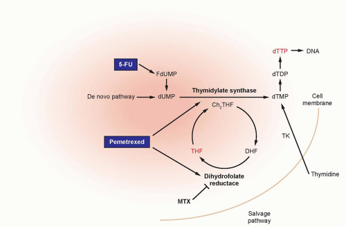
by several biochemical pathways. It is thought that 5-FU exerts its cytotoxic effects through various mechanisms, including (1) the inhibition of TS, (2) incorporation into RNA, and (3) incorporation into DNA (Fig. 19.1). In addition to these mechanisms, the genotoxic stress resulting from TS inhibition may also activate programmed cell-death pathways in susceptible cells, which leads to the induction of parental DNA fragmentation.


Figure 19.1 Antifolates and 5-fluorouracil (5-FU) sites of action. FdUMP, fluorodeoxyuridine monophosphate; dUMP, deoxyuridine monophosphate; dTTP, deoxythymidine triphosphate; dTDP, deoxyuridine diphosphate; dTMP, deoxythymidine monophosphate; TK, thymidine kinase; CH2THF, 5,10-methylenetetrahydrofolate; THF, tetrahydrofolate; DHF, dihydrofolate.


Mechanisms of Resistance

Several resistance mechanisms to 5-FU have been identified in experimental and clinical settings. Alterations in the target enzyme TS represent the most commonly described mechanism of resistance. In vitro, in vivo, and clinical studies have documented a strong correlation between the levels of TS enzyme activity/TS protein and chemosensitivity to 5-FU. In this regard, cell lines and tumors with higher levels of TS are relatively more resistant to 5-FU. Mutations in the TS protein have been identified that lead to reduced binding affinity of the 5-FU metabolite fluorodeoxyuridine monophosphate (FdUMP) to the TS protein. Reduced expression and/or diminished activity of key activating enzymes may interfere with the formation of cytotoxic 5-FU metabolites. Decreased expression of mismatch repair enzymes, such as human mutL homolog 1 (hMLH1) and human mutS homolog 2 (hMSH2), and increased expression of the catabolic enzyme dihydropyrimidine dehydrogenase (DPD) are associated with fluoropyrimidine resistance. At this time, the relative contribution of each of these mechanisms in the development of cellular resistance to 5-FU in the actual clinical setting remains unclear.


Clinical Pharmacology

5-FU is not orally administered, given its erratic bioavailability resulting from high levels of the catabolic enzyme DPD present in the gut mucosa. After intravenous bolus doses, metabolic elimination is rapid, with a half-life of 8 to 14 minutes. More than 85% of an administered dose of 5-FU is enzymatically inactivated by DPD, the rate-limiting enzyme in the catabolism of 5-FU.

A pharmacogenetic syndrome has been identified in which partial or compete deficiency in the DPD enzyme is present in 3% to 5% and 0.1% of the general population, respectively. As DPD catalyzes the rate-limiting step in the catabolic pathway of 5-FU, a deficiency of DPD can result in a clinically dangerous increase in the anabolic products of 5-FU. Unfortunately, patients with DPD deficiency do not manifest a phenotype only until they are treated with 5-FU, and in that setting, they can develop severe GI toxicity in the form of mucositis and/or diarrhea, myelosuppression, neurologic toxicity, and in rare cases, death. In patients being treated with 5-FU or any other fluoropyrimidine, it is important to consider DPD deficiency in patients who present with excessive, severe toxicity.12 It is now increasingly appreciated that DPD mutations are unable to account for all of the observed cases of excessive 5-FU toxicity, because up to 50% of patients who experience 5-FU toxicity will have no documented alterations in the DPD gene. Moreover, individuals with normal DPD enzyme activity may be diagnosed with high plasma levels of 5-FU, resulting in increased toxicity. Although DPD enzyme activity can be assayed from peripheral blood mononuclear cells in a specialized laboratory, routine phenotypic and genotypic screenings for DPD deficiency prior to 5-FU therapy are not yet available.


Biomodulation of 5-FU

Significant efforts have focused on enhancing the antitumor activity of 5-FU through biochemical modulation in which 5-FU is combined with various agents, including leucovorin, MTX, N-phosphonacetyl-l-aspartic acid, interferon-α, interferon-γ, and
a whole host of other agents.13 For the past 20 to 25 years, the reduced folate LV has been the main biochemical modulator of 5-FU. An alternative approach has been to alter the schedule of 5-FU administration. Given the S-phase specificity of this agent, prolonged exposure of tumor cells to 5-FU would increase the fraction of cells being exposed to the drug. Overall response rates are significantly higher in patients treated with infusional schedules of 5-FU than in those treated with bolus 5-FU, and this improvement in response rate has translated into an improved progression-free survival. Moreover, the overall safety profile is improved with infusional regimens. A hybrid schedule of bolus and infusional 5-FU was originally developed in France, and this regimen has shown superior clinical activity compared with bolus 5-FU schedules. This hybrid schedule has now been simplified by using only the 46-hour infusion of 5-FU and completely eliminating the 5-FU bolus doses.


TABLE 19.2 Toxicities of Different Forms of 5-FU


Route


Schedule


Dose


DLT


IV


Daily × 5, bolus


400-500 (mg/m2/d)


image BM


D


M


IV


Weekly bolus


450-500 (mg/m2/d)


image BM


IV


Daily × 5, CI


750-1,000 (mg/m2/d)


M


D


IV


PCI


200-400 (mg/m2/d)


M


HFS


HAI


Daily × 14-21, CI


750-1,000 (mg/m2/d)


M


D


IP


32-120 hr


5 nM


M


D


Oral (Xeloda)


14-21 d


2,000-2,500 (mg/m2/d)


HFS


DLT, dose limiting toxicity; IV, intravenous; BM, bone marrow; D, diarrhea; M, mucositis; CI, continuous infusion; PCI, protracted continuous infusion; HFS, hand-foot syndrome; HAI, hepatic artery infusion; IP, intraperitoneal.

Only gold members can continue reading. Log In or Register to continue

Stay updated, free articles. Join our Telegram channel

Jun 28, 2016 | Posted by in ONCOLOGY | Comments Off on Antimetabolites

Full access? Get Clinical Tree

Get Clinical Tree app for offline access