Nervousness
Heat intolerance
Insomnia
Anxiety
Hyperhidrosis
Weight loss
Fatigue
Muscle weakness
Decreased menstrual flow
Palpitations
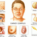
Signs
Tachycardia
Ophthalmopathy
Dermopathy (pretibial myxedema)
Proximal muscle weakness
Diffuse goiter (+ /- bruit)
Fine tremor
Spooning of nails
Graves’ disease is characterized by suppressed TSH level and an elevated serum-free T4. The suppressed TSH level helps exclude other conditions in which the serum T4 may be elevated, such as estrogen therapy, nonthyroidal illness, TSH-secreting pituitary adenoma, or central thyroid hormone resistance [7].
Once the diagnosis of thyrotoxicosis is established, radionuclide scintigraphy may be useful in differentiating Graves’ disease from other causes of hyperthyroidism. In Graves’ disease, the thyroid gland is diffusely enlarged with intense, diffuse radiotracer uptake often as high as 80 % in 24 h. The uptake is homogeneous whereas in TMG there are areas of both increased and decreased uptake. The overall uptake in TMG is not as avid as in Graves’ disease. In toxic adenoma, there is focal uptake in a single nodule, often with suppression of the remaining normal glandular tissue (Table 2) .
Table 2
Radionuclide scanning and hyperthyroidism
Scan findings |
|---|
Graves’ disease intense diffuse uptake |
Toxic adenoma focal uptake with suppressed adjacent tissue |
Toxic multinodular goiter areas of patchy uptake |
On ultrasound, patients with Graves’ disease have a diffusely enlarged thyroid gland, with a smooth but lobular surface contour. The thyroid gland may range from being isoechoic to diffusely hypoechoic, and is typically homogeneous and with increased vascularity often referred to as “the thyroid inferno” [8].
The treatment of Graves’ disease is directed at decreasing the B-adrenergic symptoms by administering a B-blocking drug and inhibiting the synthesis and release of thyroid hormone thus reversing the catabolic effects. B-blocking agents such as propranolol and atenolol can quickly reduce symptoms of palpitations, nervousness, sweating, and tremor. Inhibition of thyroid hormone synthesis and release can be achieved by thionamide drugs, radioiodine (RAI) therapy, and surgery (Table 3) .
Table 3
Hyperthyroidism and treatment options
Thionamides | RAI | Surgery | |
|---|---|---|---|
Graves’ disease | a | a | a |
Toxic adenoma | – | c | a |
Toxic multinodular goiter | – | b | a |
The two most common thionamide drugs are methimazole (Tapazole, MMI) and propylthiouracil (PTU). Both agents are equally effective. MMI has a greater potency and longer biological half-life, whereas PTU has the theoretical advantage of a more rapid fall in serum T3 levels because of its property of inhibiting peripheral conversion of T4 to T3 [9]. Biochemical euthyroidism is usually established within 6–8 weeks and doses are adjusted accordingly. The drugs are well tolerated with minimal side effects. Approximately 20 % of patients are allergic to both agents, necessitating the use of RAI or surgery. The most serious side effect of the drugs is agranulocytosis, which is rare and can occur in 0.2–0.5 % of patients, and can be fatal. However, elevations in liver enzymes may also necessitate cessation of thionamides [10, 11] .
Complete remission rates with thionamides vary considerably; however, studies have shown up to 75 % remission rate with high-dose therapy. Most relapses following cessation of thionamide therapy occur shortly after the drugs are discontinued. In some cases, relapses can occur even several years later; therefore, these patients need to be followed closely for extended periods of time. Patients who fail thionamide therapy will require definitive therapy, either RAI or total thyroidectomy.
In the USA, RAI therapy is the most common choice of therapy for Graves’ disease [12]. Its benefits include general effectiveness, relatively low expense, and minimal side effects. However, RAI therapy may take months to have an effect. The most common side effect is dry mouth which may be temporary and in rare cases permanent. The most considerable side effect is hypothyroidism which is actually the intended goal of therapy. The risk of secondary malignancies from RAI therapy for Graves’ disease is very small and usually not considered significant for adult patients [13, 14]. The risk of developing deleterious genetic, carcinogenic, teratogenic, and reproductive effects from RAI therapy for Graves’ disease is negligible [15–17]. Contraindications for RAI therapy include pediatric age, pregnancy, and nursing mothers. Patients treated with RAI therapy should avoid becoming pregnant for at least 6 months [15] .
Surgical management of Graves’ disease is becoming more common in the USA [18]. Advantages to surgery include its high success rate, rapid onset of effect, and relative safety. Both thionamide and RAI therapy can take weeks to months to achieve euthyroidism. Thyroidectomy offers immediate therapeutic response. The risk of surgery, including hypoparathyroidism and vocal cord paralysis, is very low in high-volume surgeons [18, 19]. Unlike the high relapse rate after discontinuation of thionamides, surgery reliably provides a cure with only a small chance of recurrent hyperthyroidism.
Patients who would certainly benefit from surgery are those with large goiters likely to be resistant to RAI, patients with compressive symptoms, and those with coincidental thyroid nodules concerning for malignancy. Approximately 13 % of patients with Graves’ disease have thyroid nodules suspicious for carcinoma [20]. Studies have shown that the incidence of malignancy for a solid, cold nodule is the same as for a patient without the disease, 15–20 %. Some studies suggest that thyroid carcinoma in patients with Graves’ disease is more likely to be aggressive, with increased lymph node metastasis and local invasion [21, 22]. Multiple other studies report no difference in the prognosis of carcinoma for patients with or without Graves’ disease .
Other indications for surgical management of Graves’ disease include medically noncompliant patients, thyroid storm unresponsive to medical therapy, amiodarone-induced thyrotoxicosis, and thyroid-associated ophthalmopathy (TAO). TAO can be seen in up to 50 % of patients with Graves’ disease [23]. Some studies have shown that total thyroid ablation by surgery or RAI decreases initiation and halts progression of TAO [24–26]. Studies have also reported that RAI may worsen TAO. Of the two ablative therapies, surgery appears to be more effective than RAI .
Preoperative preparation is recommended to avoid intraoperative or postoperative thyroid storm. Many different regimens have been utilized. Most regimens consist of a combination of thionamide therapy, iodine (SSKI, Lugol’s solution), and/or B-blocking agent. The choice of operation also varies based on surgeon preference and should be individualized for each patient. The three main operations are total thyroidectomy, bilateral subtotal thyroidectomy, and total lobectomy with contralateral subtotal lobectomy. The goal is to avoid hypoparathyroidism and injury to the recurrent laryngeal nerves, and at the same time minimize the chance of recurrent hyperthyroidism. Most authors recommend total thyroidectomy when possible. The other options leave remnant tissue and are associated with an increased rate of recurrent hyperthyroidism. After surgery , most patients are rendered hypothyroid and life-long thyroid hormone replacement therapy should be anticipated .
Toxic Nodular Goiter
The term toxic nodular goiter refers to two entities: toxic adenoma, implying a single lesion, and toxic multinodular goiter (TMG), in which more than one hyperfunctioning nodule exists . Toxic nodular goiter was first described as a type of thyrotoxicosis clinically distinct from Graves’ disease by Henry Plummer in 1913. Often the term Plummer’s disease is used to describe thyrotoxicosis resulting from either a single nodule or multiple autonomous nodules .
Toxic Adenoma
Toxic adenomas are discrete, solitary nodules that may occur at any age. They are most common in the third to fourth decade of life and they are rare in children [27, 28] . Toxic adenoma synthesizes and secretes thyroid hormone independent of TSH control. This results in TSH suppression, with increased T3 and T4 levels. Usually the nodule is the only functioning tissue with the extranodular tissue becoming dormant. This is highlighted in the hotspots seen with radionuclide scanning (Table 2). Toxic adenomas seem to be more prevalent in regions of iodine deficiency and recently it has been shown that a mutation of the gene for the TSHR may be associated with their development. This mutation results in the constitutive activation of the cyclic adenosine monophosphate (cAMP) cascade, causing hypersecretion of thyroid hormone and tissue growth [29–32].
Most toxic adenomas are follicular adenomas with carcinoma being very rare. Hyperthyroidism usually does not occur until the nodule is 2.5–3.0 cm in diameter. The symptoms and signs of hyperthyroidism are milder than in Graves’ disease and do not include ophthalmopathy and dermopathy. The diagnosis is usually established by a suppressed TSH and elevated free T3 or free T4 levels. In subclinical thyrotoxicosis, serum levels of T3 and T4 may be normal. Confirmation of the diagnosis may require radionuclide scanning, showing concentration of RAI in the nodule with inhibition of RAI uptake in surrounding thyroid tissue. Fine-needle aspiration biopsy of toxic adenomas is not recommended. They are rarely malignant and more importantly, cytologic features of toxic adenomas can be misleading. These lesions often exhibit cellular atypia suggestive of a follicular neoplasm or well differentiated thyroid cancer [33] .
The treatment for toxic adenomas is either ablation or surgery (Table 3). Unlike Graves’ disease , there is no role for thionamides in the management of toxic adenomas. Long-lasting remission rarely occurs with thionamide therapy and after therapy is stopped, the chance of recurrent hyperthyroidism is high. Radioiodine therapy can be effective but the doses required are usually higher than those used in Graves’ disease. Also, complete nodule regression with RAI therapy is not common and continued surveillance is necessary. This makes RAI therapy less desirable than surgery. Sclerosing agents such as ethanol have been shown to be effective in several small series [34, 35]. However, multiple injections are often needed (3–13) and exacerbation of hyperthyroidism and temporary vocal cord paralysis have been reported with ethanol injection [36, 37] .
Surgery is commonly employed in the treatment of toxic adenomas and is usually the treatment of choice. Unilateral thyroid lobectomy is the preferred procedure by most authors. It is very effective with low risk of hypothyroidism since the contralateral lobe is not removed. Surgery also provides tissue for pathologic diagnosis in the rare cases of suspected carcinoma .
Toxic Multinodular Goiter
The prevalence of TMG is significantly higher in areas of endemic goiter and iodine deficiency. In areas where iodine repletion has occurred, the prevalence of TMG has decreased [38]. A genetic predisposition and female gender also seem to play a role in the development of TMG .
TMG seems to evolve over many years from sporadic, diffuse goiter to the development of functional autonomy and eventual clinical thyrotoxicosis. The pathogenesis of TMG likely involves iodine deficiency which leads to a decreased production of thyroid hormone, resulting in an increase in TSH secretion, promoting goiter formation [39, 40]. Multiple nodules develop which in time become autonomous. Autonomous areas eventually grow large enough to secrete increased amounts of thyroid hormone and suppress TSH. Hyperthyroidism can be precipitated in nontoxic multinodular goiter both with autonomy and without autonomy and in TMG by iodides (i.e., intravenous (IV) contrast media) [41]. This is referred to as the Jod-Basedow phenomenon.
Due to the many years it takes to develop, TMG generally occurs in older persons. The hyperthyroidism tends to be insidious in onset, may be mild to severe, and is unaccompanied by the infiltrative ophthalmopathy and dermopathy of Graves’ disease. In older patients, the hyperthyroidism may be masked and the patient may present with cardiac findings such as atrial fibrillation, tachycardia, congestive heart failure, angina, weight loss, anxiety, insomnia, or muscle wasting. Patients with TMG often have no thyromegaly on clinical examination .
The diagnosis of TMG is a clinical one, based on physical examination and laboratory confirmation. Serum levels of free T3 and free T4 are elevated with a suppressed TSH. Radionuclide scanning reveals a multinodular gland with areas of increased patchy uptake (Table 2).
The principal treatment options for TMG include RAI therapy or surgery (Table 3). Since remission does not occur with TMG, the long-term use of thionamides is not indicated unless there are contraindications to the use of RAI therapy or surgery . Patients with TMG often require multiple doses of RAI to control hyperthyroidism because of the larger gland size and lower uptake of RAI, when compared to patients with Graves’ disease. Although RAI treats the hyperthyroidism, studies show that it does not significantly reduce goiter size, compressive symptoms, or substernal extension, because TMG contains areas of fibrosis, calcifications, and nonfunctioning nodules [42, 43] .
Surgery is usually recommended in younger, healthier patients with large goiters and/or compressive symptoms. Either bilateral subtotal thyroidectomy or total thyroidectomy is the preferred operation. Bilateral subtotal thyroidectomy has the potential advantage of decreased hypoparathyroidism, decreased vocal cord paralysis, and decreased hypothyroidism. However, the incidence of recurrent hyperthyroidism is greater than in total thyroidectomy patients. Total thyroidectomy results in near-zero recurrence but nearly all patients are rendered hypothyroid, requiring thyroid hormone supplementation. Total thyroidectomy for patients with TMG is a safe operation in experienced hands with low rates of hypoparathyroidism and vocal cord paralysis [44] .
Thyroiditis
Thyroiditis , infiltration of the thyroid gland with inflammatory cells, may be seen in a diverse group of autoimmune, inflammatory, and infectious processes. It comprises a diverse group of disorders that are among the most common endocrine abnormalities encountered. The diagnosis of thyroiditis is based on the clinical presentation and laboratory analysis of thyroid function. Thyroiditis can be classified as (1) chronic, which includes Hashimoto’s thyroiditis and Riedel struma, (2) subacute, which includes lymphocytic and granulomatous, and (3) acute suppurative, which is rare (Table 4) .
Table 4
Different types of thyroiditis
Hashimoto’s thyroiditis (chronic lymphocytic thyroiditis) |
Subacute (painless) lymphocytic thyroiditis Sporadic silent thyroiditis Postpartum thyroiditis |
Subacute (painful) granulomatous thyroiditis (de Quervain’s thyroiditis, giant cell thyroiditis) |
Acute suppurative thyroiditis |
Riedel struma (Riedel’s thyroiditis, invasive fibrous thyroiditis) |
Hashimoto’s Thyroiditis
Hashimoto’s thyroiditis, also called chronic lymphocytic thyroiditis, is the prototypical autoimmune thyroiditis . Hashimoto’s is the most common cause of goiter and hypothyroidism in the USA and affects approximately 2 % of the general population [45]. Hakaru Hashimoto first described this disorder in 1912 and termed it “struma lymphomatosa.”
Pathologically, the thyroid gland is initially enlarged and has lymphocytic and plasma-cell infiltration, follicular cell atrophy, and interlobular fibrosis, eventually leading to a shrunken fibrotic gland [46]. The normal follicular cells are altered and often replaced with pink oxyphilic or Hurthle cells. Classically, Hashimoto’s thyroiditis occurs as a painless diffuse goiter in young to middle-aged women in their third and fourth decades and is frequently associated with asymptomatic hypothyroidism [47] . Hashimoto’s thyroiditis has also been reported to occur with increased frequency in patients with other autoimmune disorders such as lupus, Graves’ disease , and pernicious anemia [48].
The hallmarks of this disorder are high circulating titers of antibodies to thyroid peroxidase (90 % of patients) and thyroglobulin (20–50 % of patients). Antibodies to the TSHR have also been identified. The inciting event that triggers the development of antithyroid antibodies remains unclear. There does appear to be a genetic predisposition, with reported associations with human leukocyte antigen (HLA)-DR3, HLA-DR5, and HLA-B8 [49–51]. Viral etiologies and smoking have also been implicated. Although the exact pathogenesis is not known, it is clear that thyroid autoimmunity drives the lymphocytic collection and is responsible for thyroid epithelial cell damage. Progressive, immune-mediated thyroid cell damage leads to goiter formation and thyroid gland failure .
The clinical presentation varies, depending on the stage at the time of presentation. The patient may be completely asymptomatic or present with hypothyroid symptoms. Physical examination typically reveals a firm, bumpy, nontender goiter, often symmetric, with a palpable pyramidal lobe. Usually there are no discrete nodules. Single or dominant nodules should be evaluated and if indicated a fine-needle aspiration biopsy should be performed to exclude malignancy. Thyroid hormone levels may also vary based on time of presentation. They may be normal with a normal TSH (euthyroid), low with an elevated TSH (hypothyroid), or normal with an elevated TSH (subclinically hypothyroid). Euthyroid individuals with Hashimoto’s thyroiditis develop hypothyroidism at a rate of approximately 5 % per year [52]. Mild thyrotoxicosis (“Hashitoxicosis”) has been reported to be the initial presentation in up to 5 % of patients with Hashimoto’s thyroiditis [53] . The clinical course for Hashimoto’s thyroiditis is variable. Up to 50 % of patients can become subclinically hypothyroid and 5–40 % can become clinically hypothyroid, emphasizing the importance of following thyroid function tests in these patients [54].
Stay updated, free articles. Join our Telegram channel

Full access? Get Clinical Tree


