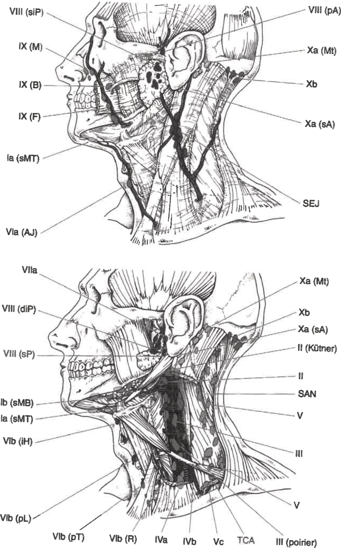
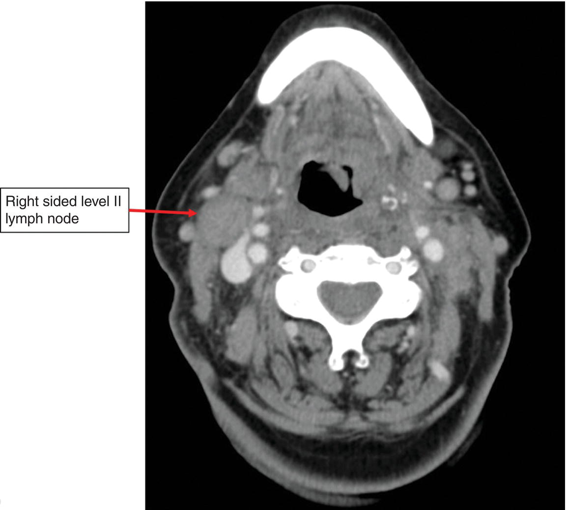
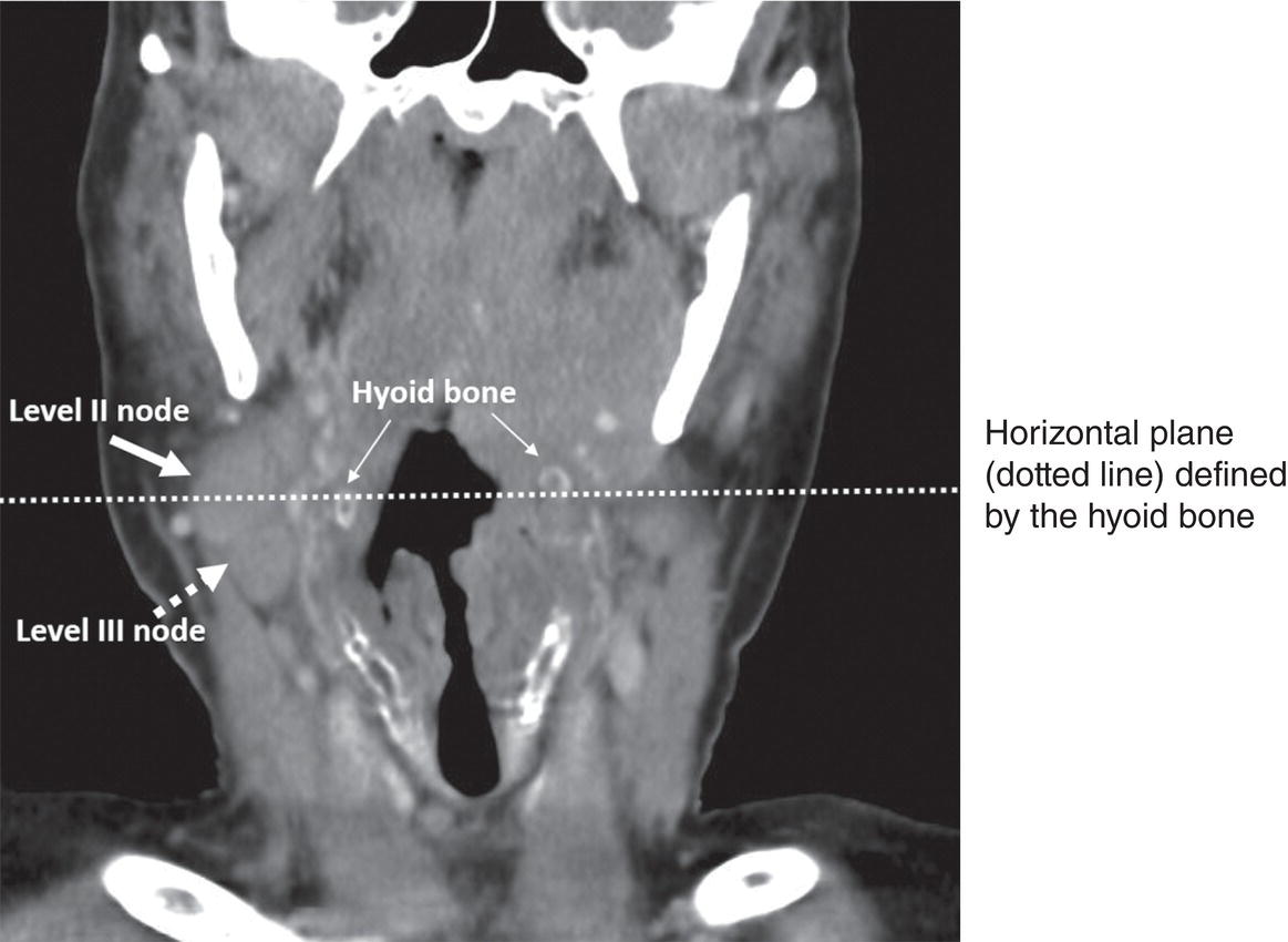
The following sites are included: Carcinomas arising in the minor salivary glands of the upper aerodigestive tract are classified according to the rules for tumours of their anatomical site of origin, e.g., oral cavity. The definitions of the N categories for all head and neck sites except p‐16 positive oropharynx, nasopharynx, mucosal malignant melanoma of the upper aerodigestive tract and thyroid are the same. Midline nodes are considered ipsilateral nodes except in the thyroid. The status of the regional lymph nodes in head and neck cancer is of considerable prognostic importance. In addition, it is helpful to subdivide the lymph nodes and possible metastasis into specific anatomical subsites and to group these lymph nodes into levels. A consensus guideline from DAHANCA, EORTC, HKNPCSG, NCIC CTG, NCRI, RTOG and TROG has been published and the nodal groups are listed below. However, a number of different classifications exist that use variable level numbers and therefore we recommend the levels be named rather than referred to by number to limit any confusion, although the levels used in the consensus document are given.1 In the consensus classification, the retropharyngeal nodes are classified as Level VII, but in the classification used by the AJCC, Level VII described the upper mediastinal nodes. Source: Modified from Lengele B et al., Radiothor Oncol, 2007; 85(1): 146–155. Fig. 2 Axial CT scan showing enlarged right upper jugular (deep cervical) Level II lymph node measuring 2.5 cm in greatest dimension. Fig. 3 Coronal CT scan showing the same enlarged right upper jugular (deep cervical) Level II lymph node measuring 2.5 cm in greatest dimension, but also an enlarged right medial jugular (deep cervical) Level III lymph node measuring 1.5 cm in greatest dimension. Horizontal plane (dotted line) delineated by the hyoid bone that defines Level II nodes superiorly from Level III nodes inferiorly is marked. This is classified as cN2b: metastasis in multiple ipsilateral lymph nodes, none more than 6 cm in greatest dimension without extranodal extension. The lymph node groups are defined as follows Lymph nodes within the triangular boundary of the anterior belly of the digastric muscle and the hyoid bone. Lymph nodes within the boundaries of the anterior and posterior bellies of the digastric muscle and the body of the mandible. Lymph nodes located around the upper third of the internal jugular vein and adjacent spinal accessory nerve, extending from the hyoid bone (clinical landmark) to the skull base. The posterior boundary is the posterior border of the sternocleidomastoid muscle, and the anterior boundary is the lateral border of the sternohyoid muscle. This group includes the jugulodigastric node, which is the most cranial jugular node. Lymph nodes located around the middle third of the internal jugular vein, extending from the carotid bifurcation superiorly to the omohyoid muscle (surgical landmark) or cricothyroid notch (clinical landmark) inferiorly. The posterior boundary is the posterior border of the sternocleidomastoid muscle, and the anterior boundary is the lateral border of the sternohyoid muscle. This group includes the jugulo‐omohyoid lymph node located between the omohyoid muscle and the internal jugular vein. Lymph nodes located around the lower third of the internal jugular vein, extending from the omohyoid muscle superiorly to the clavicle inferiorly. The posterior boundary is the posterior border of the sternocleidomastoid muscle, and the anterior boundary is the lateral border of the sternohyoid muscle.
HEAD AND NECK TUMOURS
Introductory Notes
Regional Lymph Nodes (Figs. 1, 2, 3)
Related posts:


Stay updated, free articles. Join our Telegram channel

Full access? Get Clinical Tree


