Aging of the Respiratory System: Introduction
This chapter first reviews the changes in lung function with aging that are known to occur in healthy persons (normal, never smokers). Included are the major categories of pulmonary function tests: static lung volumes, maximal expiratory flow, lung mechanics, and gas exchange, plus bronchodilator (BD) response and nonspecific airway reactivity. Table 82-1 summarizes these changes. The chapter then adds a discussion of how lung function tests maybe used by a clinician to assist in the differential diagnosis of asthma and chronic obstructive pulmonary disease (COPD) in an elderly patient, and to objectively measure the efficacy of asthma and COPD therapy (see Chapter 83 for additional information on this topic).
Lower maximal expiratory flows: FEV1, FEV1/FVC, FEF 75% |
Increased FRC and RV, lower VC, but stable TLC |
Lower diffusing capacity (oxygen uptake) |
Lower PO2 and SpO2 as a consequence of V/Q mismatch (but no change in PCO2) |
Lower respiratory muscle strength and endurance |
Stiffer chest wall (less compliant) |
Increased lung tissue compliance (loss of lung recoil) |
Reduced respiratory drive (for hypoxia, hypercarbia, and resistive loads) |
Increased airway reactivity (but no change in bronchodilator responsiveness) |
Lung Volumes
Total lung capacity (TLC) is the volume of air within the respiratory system when a subject makes a maximal voluntary inspiratory effort (and the air seen in the lungs on a chest x-ray). It is determined by the balance of forces between the maximally activated inspiratory muscles and the elastic recoil of the lung and chest wall. A decrease in TLC and other static lung volumes is called restriction.
The elastic recoil of lung tissue decreases with aging (just as the skin becomes less elastic), making the lungs easier to expand during a deep breath toward TLC. This reduction of elastic recoil tends to increase TLC; however, the chest wall (rib cage) becomes stiffer with aging, so that a maximal inspiratory effort is not able to achieve a higher lung volume even though the lungs themselves have become easier to expand. Thus, TLC normally remains stable throughout the aging process.
The volume of air remaining in the lungs when subjects have exhaled as much air as possible is called residual volume (RV). Because lung elastic recoil decreases as a consequence of normal aging, the RV and RV/TLC increase from young adulthood to older age. An abnormally high RV/TLC is called hyperinflation, can often be seen on a chest x-ray, and occurs with both asthma and COPD.
Vital capacity (VC) is the difference between the absolute lung volume at TLC and at RV, and the amount of air that a person can slowly exhale after inhaling maximally. Because TLC is relatively constant while RV increases with age, the VC decreases with age. Hutchinson coined the term vital capacity over 150 years ago because it predicted the vigor and length of life of his patients. Many modern epidemiologic studies have verified his observation.
Maximal Expiratory Flow
Maximal expiratory flow, as seen during spirometry tests, varies as a function of lung volume because higher flows are possible at a higher lung volume. For forced exhalation beginning from a deep inhalation (the first phase of a spirometry maneuver), the initial (peak) flow (phase 2) is determined by the recoil of the lung and chest wall and the degree of effort expended by the patient, as well as by the speed with which the patient’s respiratory muscles can generate positive pleural pressure. Once maximal flow is achieved, maximal flow throughout the remainder of the VC (phase 3) is determined by the intrinsic properties of the lung.
Spirometry
Spirometry is the most common test of lung function, and is easily performed by 9 of 10 elderly patients. Modern office spirometers use a flow sensor, connected to a microprocessor that determines the forced expiratory volume in one second (FEV1), forced vital capacity (FVC), and FEV1/FVC from the best maneuver, as well as the predicted values. FEV1 is the most important value, representing the volume of air (in liters) exhaled during the first second. Guidelines for spirometry methods and instrument accuracy are available from the American Thoracic Society.
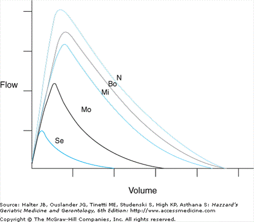
The presence of airways obstruction is determined by a low FEV1/FVC ratio and visualized on the flow–volume (F –V) curve as concavity toward the volume axis or tail at the end of the maneuver (Figure 82-1). The descending limb of the F–V curve of healthy young adults is a straight line of about 45 degrees until the end of the maneuver (shaped like a sail), corresponding to exponential emptying of the lungs. The shape of the F–V curve becomes progressively more curvilinear as a healthy adult becomes older, corresponding to decreases in expiratory flows at low lung volumes. The reduced flow at low lung volumes, even in healthy elderly persons, is a result of a decrease in the mean diameter of small airways.
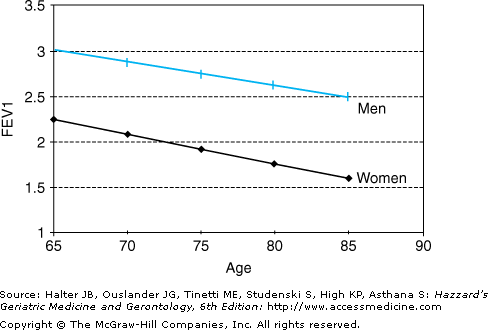
Adults normally experience a loss in FEV1 of about one-third liter per decade (Figure 82-2). The decline in FEV1 with aging is greater than the decline in FVC, so that the FEV1/FVC also declines with age. Values below 0.65 usually indicate airway obstruction in patients aged 65 years and older.
