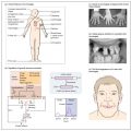
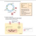
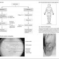
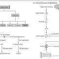
Adrenocortical insufficiency occurs as primary adrenal failure when the pathology lies in the adrenal gland, rather than secondary to failure of ACTH secretion. The symptoms may be vague, such as in the case of AD, a 22-year-old woman who presented with lethargy and fatigue. She was known to have autoimmune hypothyroidism and had been on thyroxine replacement therapy for 2 years. She now complained of increasing lethargy and intermittent abdominal pain. She became extremely unwell one day following an episode of food poisoning and was admitted to hospital as an emergency with diarrhoea, vomiting and hypotension. Examination revealed evidence of increased pigmentation of the palmar creases, over the knees and in the mouth, and hypotension with a postural fall in blood pressure of 30 mmHg. Blood tests showed her to have a blood sugar of 2.6mmol/L, Na 131 mmol/L, K 5.6 mmol/ L and urea 12.6 mmol/L. A blood sample was saved for cortisol and ACTH estimation and she was treated with intravenous and subsequently oral hydrocortisone replacement therapy. Later results showed the plasma cortisol to be 95 nmol/L in combination with an ACTH concentration of 205 ng/L. A diagnosis of Addison’s disease was made and she received advice from the endocrinology team about emergency hydrocortisone therapy, ‘sick day rules’ covering what to do in the event of intercurrent illness, and how to obtain a Medic-alert bracelet to wear continuously.
Addison’s disease. First described by Addison in 1855, the disease is caused by the destruction of adrenal tissue. The disease is usually autoimmune in origin, with the detection of adrenal autoantibodies in the plasma of 75-80% of patients, but there are numerous other causes (Table 21.1). The disease may present first as an Addisonian crisis, with fever, abdominal pain and hypotensive collapse and pigmentation of skin (Fig. 21a
Stay updated, free articles. Join our Telegram channel

Full access? Get Clinical Tree


