Adenosquamous Carcinoma
Timothy Craig Allen
Adenosquamous carcinoma is an uncommon, mixed subtype of non-small cell lung cancer that makes up approximately 2% to 5% of lung cancers.1,2,3 and 4 The incidence of adenosquamous carcinoma may be increasing, however, along with adenocarcinoma.5 Clinically, patients with adenosquamous carcinoma are usually smokers and typically exhibit symptoms similar to patients with tumors of other histologies, including cough and dyspnea, chest pain, and hemoptysis.6 Radiographically, adenosquamous carcinoma is characterized on CT scan as a solid, lobulated mass, usually peripheral, and typically exhibits heterogeneous soft tissue attenuation after intravenous injection of positive contrast medium.7,8 There may be central scarring and peripheral ground glass opacification, as well as puckering of the overlying pleura.9,10 and 11 Adenosquamous carcinomas generally exhibit earlier metastases and have a poorer prognosis than squamous cell carcinomas or adenocarcinomas.4,9,10,11,12,13,14,15 and 16 Metastatic tumors often show both adenocarcinomatous and squamous cell carcinomatous differentiation, as with the primary tumor.17
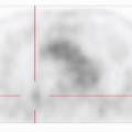
ADENOSQUAMOUS CARCINOMA, GROSS FEATURES
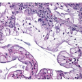
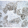
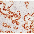
ADENOSQUAMOUS CARCINOMA, HISTOLOGY
Histologically, adenosquamous carcinoma is characterized by exhibiting areas both of adenocarcinoma and squamous cell carcinoma, each involving 10% or more of the tumor (Fig. 7-2).9,10 and 11,17 Diagnostic criteria for squamous cell carcinoma and adenocarcinoma, specifically single-cell keratinization, keratin pearls, or intercellular bridges; and acinar, tubular, or papillary structures, respectively, must be clearly and unequivocally identified in order to make the diagnosis (Figs. 7-3 and 7-4).9,10 and 11,17 Papillary and acinar patterns of adenocarcinoma may be easily recognizable; however, solid pattern adenocarcinoma with mucin formation may be more difficult to recognize, and some squamous cell carcinomas may show focal mucin positivity. In order to diagnose solid pattern adenocarcinoma, more than 5 mucin droplets per high-power field are required on mucin stain for the diagnosis of adenocarcinoma.17 The different tumor types are independently, and may be variably, differentiated. The different tumor types may be arranged separately or may be mixed with each other. Areas of large cell carcinoma may also be present in adenosquamous carcinoma. Because of the diagnostic requirement that 10% or more of each component be present in order to make the diagnosis, adenosquamous carcinoma cannot definitively be diagnosed by transbronchial biopsy or core needle biopsy.11
Stay updated, free articles. Join our Telegram channel

Full access? Get Clinical Tree


