Acquired Disorders of Red Cell, White Cell, and Platelet Production

Chapter 6 Acquired Disorders of Red Cell, White Cell, and Platelet Production


Table 6-2 Classification of Neutropenia


Congenital  
Primary Autoimmune neutropenia
  Pure white cell aplasia
  Idiopathic
  Thymoma
  Hematologic malignancies (e.g., T-LGL leukemia)
  Infections/postinfectious
  Viral
  Measles,53 mumps, roseola,54,55 rubella,56 RSV, influenza57
  Hepatitis A,58 B,58,59 and C60
  CMV,6163 EBV,6466 HIV67,68
  Parvovirus6971
  Bacterial
  Tuberculosis72,73
  Brucellosis7476
  Tularemia77
  Typhoid fever78
  Rickettsial
  Rocky Mountain spotted fever79
  Ehrlichiosis80,81
  Fungal
  Histoplasmosis82,83
  Parasitic
  Malaria,84 leishmaniasis85,86
  Autoimmune conditions (e.g., SLE87,88, RA89)
  Drugs and chemicals
  Neutropenia associated with immunodeficiency90,91
  Severe nutritional deficiencies92,93
  Neutropenia due to increased margination
  Iatrogenic (e.g., hemodialysis94,95)

CMV, Cytomegalovirus; EBV, Epstein-Barr virus; HIV, human immunodeficiency virus; RA, refractory anemia; RSV, respiratory syncytial virus; SLE, systemic lupus erythematosus; T-LGL, T-cell large granular lymphocyte.


Approach to the Diagnosis and Treatment of Acquired Pure Red Cell Aplasia, Acquired Neutropenia, and T-Cell Large Granular Lymphocyte Leukemia
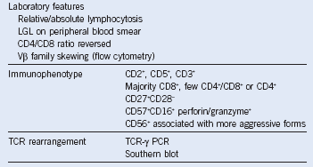

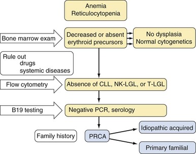
Acquired pure red cell aplasia (PRCA) is characterized by reticulocytopenia, but diagnosis is based on the morphologic absence of erythroid precursors in the bone marrow. Congenital forms of PRCA, including Diamond-Blackfan anemia, need to be distinguished when presenting in young children. In adults, primary idiopathic disease has to be differentiated from secondary forms of red cell aplasia associated with hematologic diseases such as B-cell chronic lymphocytic leukemia, myeloma, T-cell large granular lymphocyte (T-LGL) leukemia, and parvovirus B19–associated chronic reticulocytopenia or acute transient aplastic crisis. The diagnosis of parvovirus B19 infection can be made on the basis of the presence of parvovirus B19–specific immunoglobulin M and by deoxyribonucleic acid (DNA) hybridization techniques. Parvovirus B19–specific polymerase chain reaction can help rule out an ongoing infection. This diagnosis is important because therapy with intravenous immunoglobulin (IVIg) (2 g/kg IV for 5 days) can be curative. The therapy of secondary red cell aplasia includes treatment of the underlying condition. It is also important to distinguish red cell aplasia from myelodysplastic syndromes, which can be associated with erythroid hypoplasia but carry a significantly worse prognosis.


In cases with thymoma, thymectomy is the usual initial treatment approach; however, incomplete responders, nonresponders, and patients who relapse are common, necessitating the addition of immunosuppressive therapy. In patients with idiopathic PRCA, therapy includes immunosuppressive agents such as prednisone (1 mg/kg orally PO for 3 months), cyclosporine A (CsA; 5 to 10 mg/kg PO daily in divided doses for 3 months), or oral cyclophosphamide (50 to 150 mg PO daily for 3 months). Second-line therapies include antithymocyte globulin (ATG; if horse ATG, 40 mg/kg IV daily for 4 days with prednisone at 1 mg/kg, and if rabbit ATG, 2.5 to 3.5 mg/kg daily IV for 4 or 5 days with prednisone at 1 mg/kg) or rituximab (375 mg/m2 IV every week for 4 weeks).


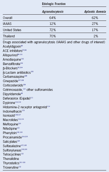
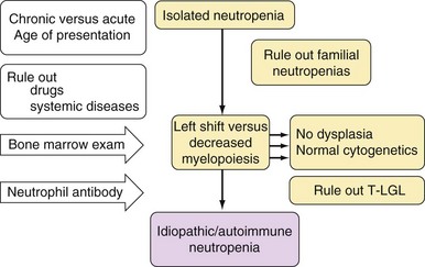
Neutropenia may be associated with severe infections, but the risk associated with neutropenia depends on its clinical context, severity, and duration. The management of neutropenia must account for its clinical presentation and the risk for possible life-threatening complications and includes supportive care, clinical monitoring, and implementation of prophylactic antibiotics and/or hematopoietic growth factors. Neutropenia in childhood may be due to congenital diseases, be associated with viral infections, or be immune mediated. These three causes are usually self-limiting. In adults, most neutropenias are secondary to other conditions, including hematologic or systemic diseases. In general, drug reactions are a very common cause of neutropenia. Idiopathic or autoimmune neutropenia as a primary disease is a diagnosis of exclusion. The pathogenesis involves T-lymphocyte/natural killer–mediated inhibition of myelopoiesis or antineutrophil antibodies. Immunosuppressive therapy may be employed and includes prednisone (1 mg/kg PO daily for 3 months), methotrexate (5 to 7.5 mg/week PO for 1 to 2 months), CsA (1 to 1.5 mg/kg PO twice a day and titrated to maintain a trough level of 250 to 400 ng/mL), IVIg (2 g/kg IV for 5 days), cyclophosphamide (50 to 100 mg PO daily for 3 to 6 months), or rituximab alone (375 mg/m2 IV every week for 4 weeks) or in conjunction with myeloid growth factors.


T-LGL leukemia is a chronic, often indolent clonal lymphoproliferation of cytotoxic T cells associated with immune-mediated cytopenias. It may be a part of a continuum of reactive cytotoxic T-cell responses ranging from polyclonal, oligoclonal, to monoclonal expansions as seen in T-LGL leukemia. Persistent antigenic drive or dysfunction of the homeostatic termination of clonal T-cell expansion may be involved, and the abnormal cytotoxic T lymphocyte (CTL) in T-LGL is not entirely autonomous. The pathophysiology of cytopenias includes cytokine effects and direct antigen-specific cytotoxicity. Most patients present with neutropenia. PRCA and pancytopenia are less common. Hemolytic anemia and pancytopenia may be the result of splenomegaly present in a significant minority of patients. B symptoms and lymphadenopathy are uncommon, and many patients remain asymptomatic. The diagnosis is established according to the presence of characteristic LGL lymphocytosis, but in some patients the LGL count may not be very high. The immunophenotype is CD3+, CD8+, CD57+, CD16+, CD56, and CD28. CD56 antigen–expressing LGL may be characterized by a more aggressive course. Some cases may coexpress CD4 and CD8. The diagnosis includes detection of T-cell receptor rearrangement. In most instances, expansion of the involved Vβ family may be detected using Vβ flow cytometric clonotyping. T-LGL is often associated with autoimmune diseases, including rheumatoid arthritis and Felty syndrome. T-LGL can accompany myelodysplasia and, in rare instances, aplastic anemia or paroxysmal nocturnal hemoglobinuria. Reactive, often viral infection–associated, CTL proliferation may be difficult to document. Asymptomatic cases are monitored, and development of systemic symptoms or symptomatic cytopenias may prompt therapy. Current treatments include immunosuppressive agents such as prednisone, CsA (1 to 1.5 mg/kg PO twice a day then adjusted to maintain a trough level of 250 to 400 ng/mL for 8 to 10 weeks), oral methotrexate (7.5 mg/m2

Stay updated, free articles. Join our Telegram channel

Jun 12, 2016 | Posted by in HEMATOLOGY | Comments Off on Acquired Disorders of Red Cell, White Cell, and Platelet Production

Full access? Get Clinical Tree

Get Clinical Tree app for offline access