The classification applies only to adenocarcinomas. Transitional cell carcinoma of the prostate is classified as a urethral tumour (see Urethra). There should be histological confirmation of the disease. The regional lymph nodes are the nodes of the true pelvis, which essentially are the pelvic nodes below the bifurcation of the common iliac arteries. Laterality does not affect the N classification. Notes 1 Tumour found in one or both lobes by needle biopsy, but not palpable, is classified as T1c. 2 Invasion into the prostatic apex or into (but not beyond) the prostatic capsule is not classified as T3, but as T2. Note Note * When more than one site of metastasis is present, the most advanced category is used. pM1c is the most advanced category. The pT and pN categories correspond to the T and N categories. However, there is no pT1 category because there is insufficient tissue to assess the highest pT category. There are no subcategories of pT2. Note
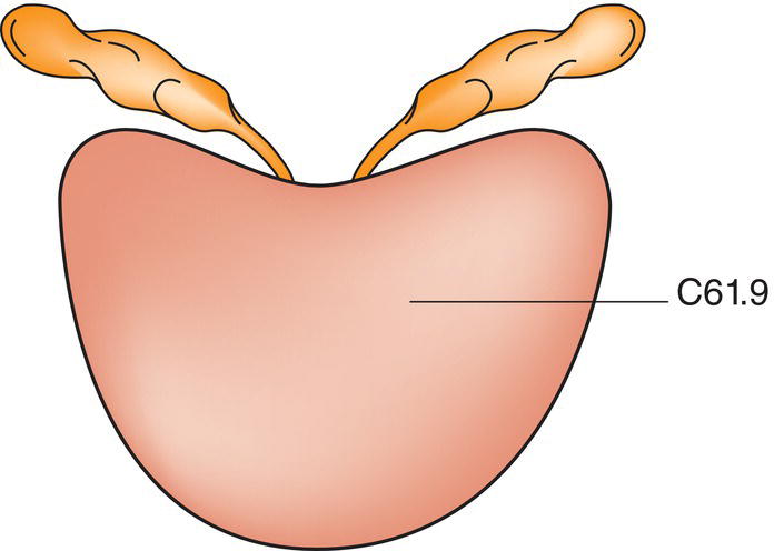
PROSTATE (ICD‐O‐3 C61) (FIG. 477, SEE ALSO FIG . 524)
Rules for Classification
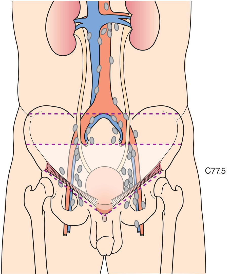
Regional Lymph Nodes (Fig. 478)
TNM Clinical Classification
T – Primary Tumour
TX
Primary tumour cannot be assessed
T0
No evidence of primary tumour
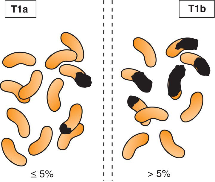
T1
Clinically inapparent tumour that is not palpable (Fig. 479)
T1a
Tumour incidental histological finding in 5% or less of tissue resected
T1b
Tumour incidental histological finding in more than 5% of tissue resected
T1c
Tumour identified by needle biopsy (e.g., because of elevated PSA)
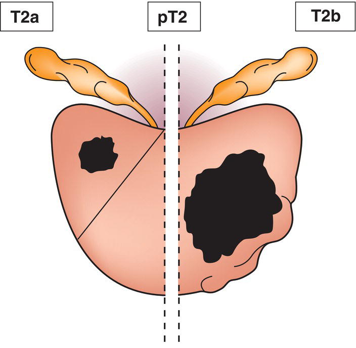
T2
Tumour that is palpable and confined within prostate1
T2a
Tumour involves one half of one lobe or less (Fig. 480)
T2b
Tumour involves more than half of one lobe, but not both lobes (Fig. 480)
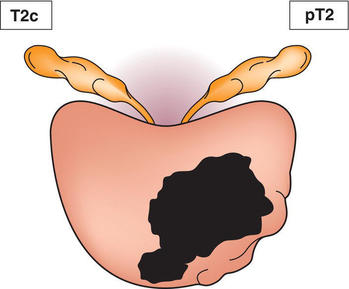
T2c
Tumour involves both lobes (Fig. 481)
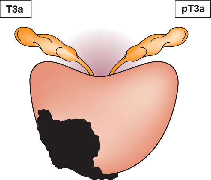
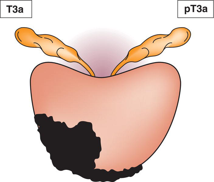
T3
Tumour extends through the prostatic capsule2
T3a
Extraprostatic extension (unilateral or bilateral) including microscopic bladder neck involvement (Figs. 482, 483)
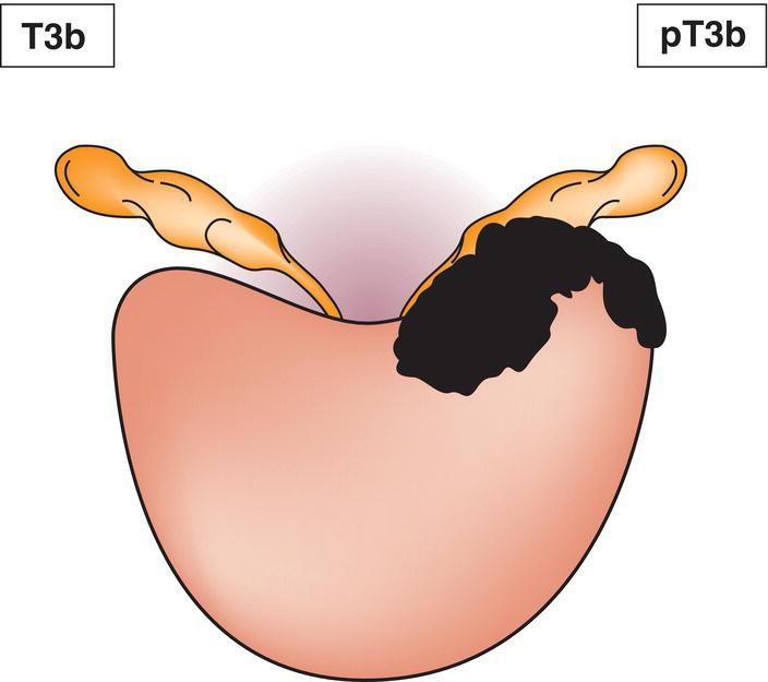
T3b
Tumour invades seminal vesicle(s) (Fig. 484)
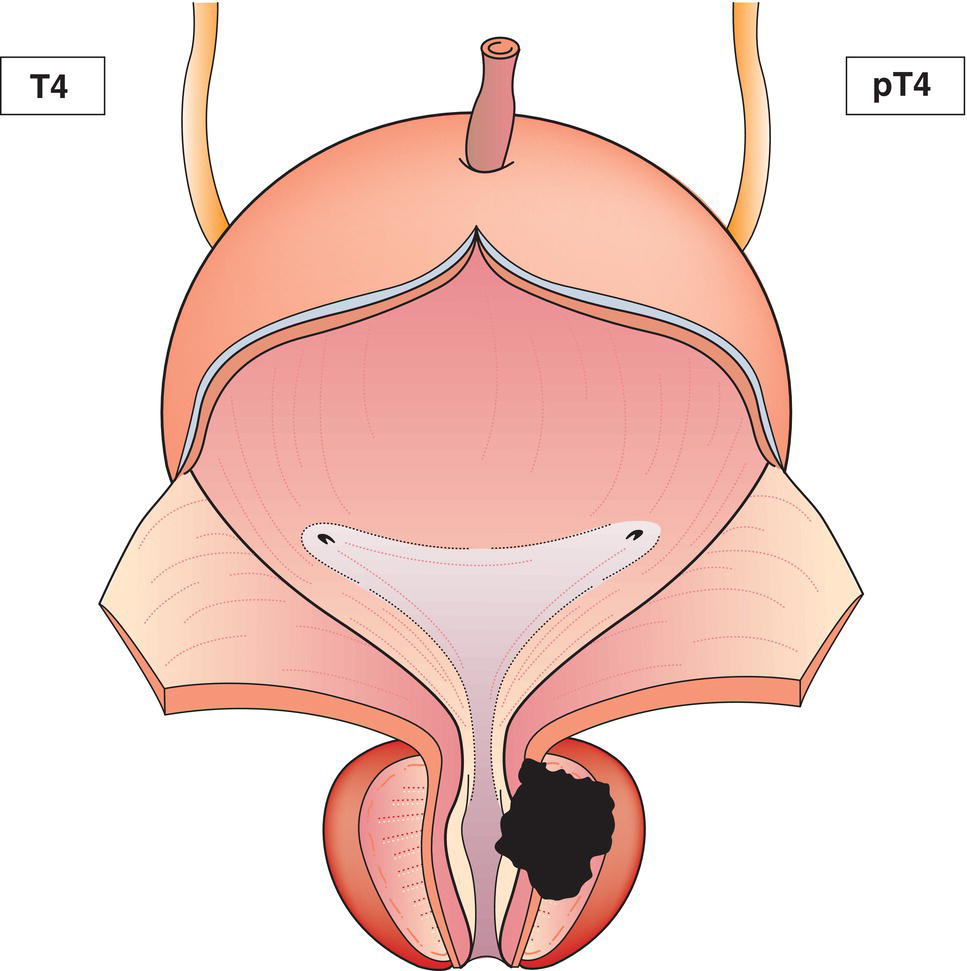
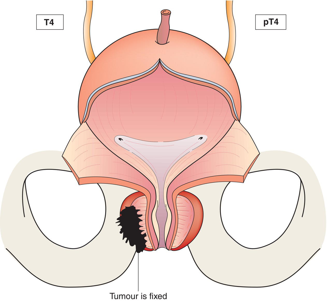
T4
Tumour is fixed or invades adjacent structures other than seminal vesicles: bladder neck, external sphincter, rectum, levator muscles, and/or pelvic wall (Figs. 485, 486)
N – Regional Lymph Nodes
NX
Regional lymph nodes cannot be assessed
N0
No regional lymph node metastasis
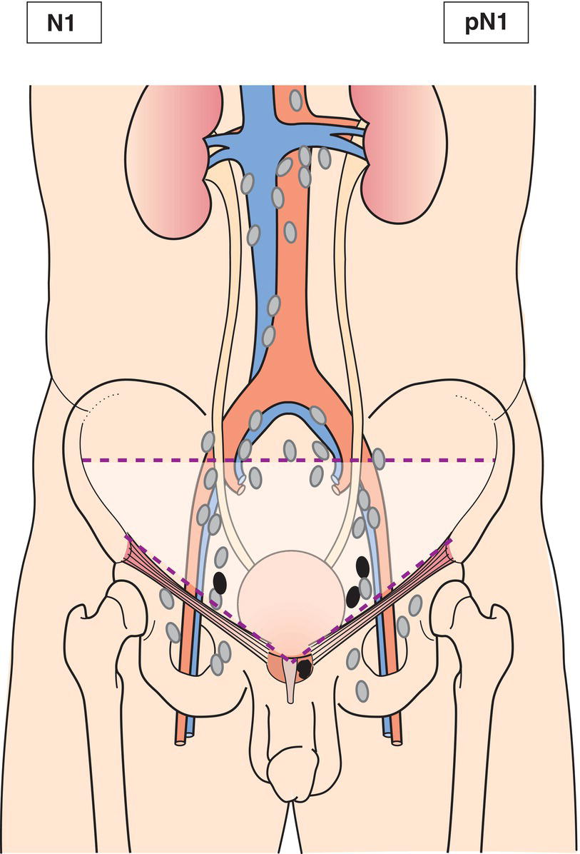
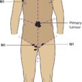
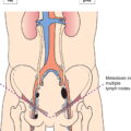
N1
Regional lymph node metastasis (Figs. 487, 488)
Metastasis no larger than 0.2 cm can be designated pN1mi.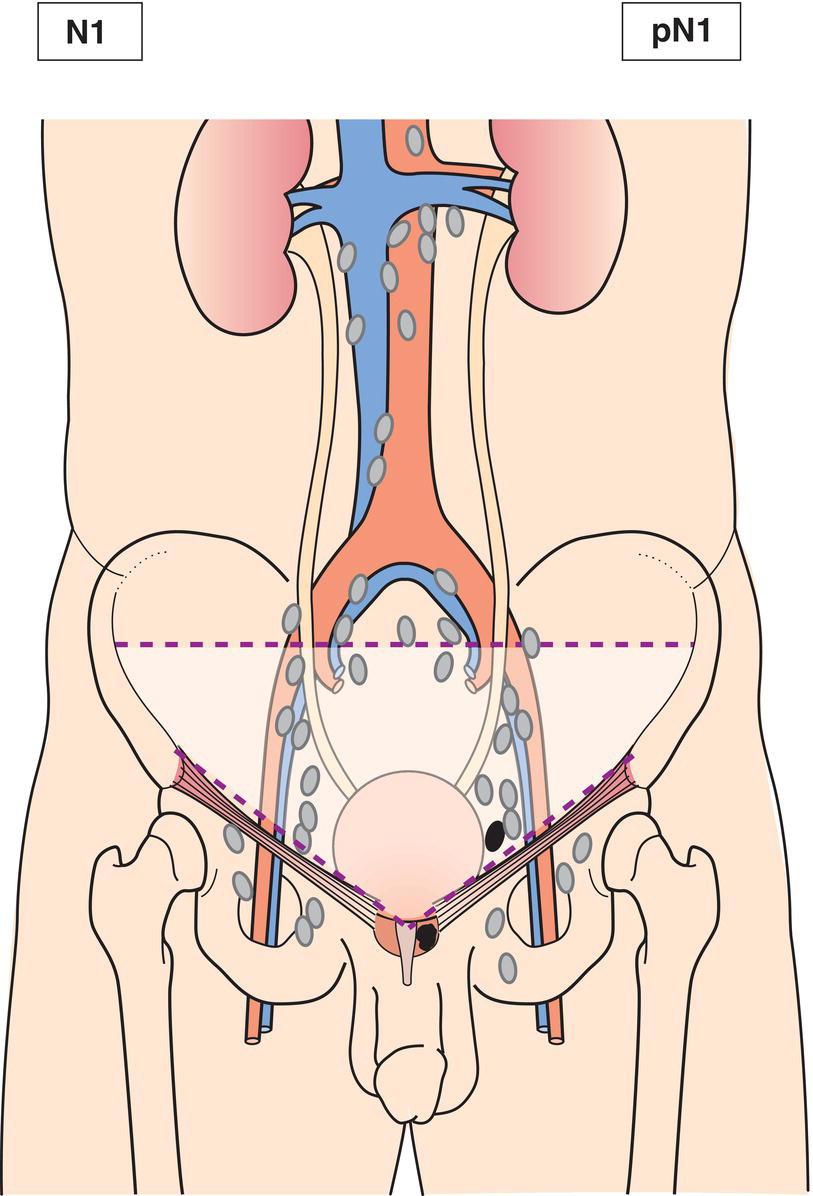
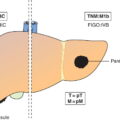
M – Distant Metastasis*
M0
No distant metastasis
M1
Distant metastasis
M1a
Non‐regional lymph node(s)
M1b
Bone(s)
M1c
Other site(s)
pTNM Pathological Classification
pM1
Distant metastasis microscopically confirmed
pM0 and pMX are not valid categories.
Summary
Related posts:


Stay updated, free articles. Join our Telegram channel

Full access? Get Clinical Tree


