9 Early Supraglottic Cancer: Horizontal Supraglottic Laryngectomy
Abstract
There is no consensus about the best treatment for supraglottic squamous cell carcinoma because we do not have randomized controlled trials comparing the different treatments. Five accepted surgical and nonsurgical oncological treatments have been currently established: standard horizontal supraglottic laryngectomy, supraglottic microsurgery, transoral robotic surgery, radiotherapy alone, and radiotherapy in combination with chemotherapy. Current treatment guidelines indicate, for early cancer, a single modality of treatment either by radiotherapy or by conservative surgery. The decision between surgery and radiotherapy should consider the patient’s choice (accept tracheostomy and feeding tube, even if temporarily), expectation regarding vocal outcome, and general clinical conditions. Based on a clinical case, this chapter discusses the rational decision-making process for an early primary supraglottic cancer, considering the clinical, radiological, and natural history of head and neck squamous cell carcinoma. In addition, we provide a step-by-step description of the horizontal standard supraglottic laryngectomy.
9.1 Case Report
A 49-year-old Caucasian male toolmaker presented with nonspecific sore throat and dysphagia for 3 months, not associated with comorbidities. His social history was positive for heavy alcohol drinking and smoking 64 packs of cigarettes per year.
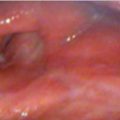
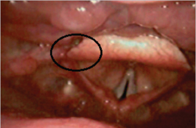
The Eastern Cooperative Oncology Group (ECOG) performance status was zero or compared as a healthy person. A nasolaryngofibroscopic evaluation revealed an ulcerative lesion of the epiglottis with minimal extension to the vallecula and left aryepiglottic fold (the anatomical position of the epiglottis impaired the visualization of the lesion). The true vocal cords were mobile and mild edema is shown in Fig. 9‑1 .
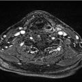
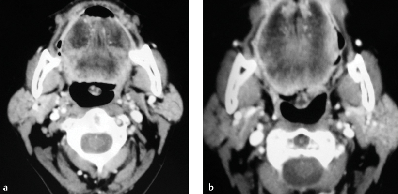
Computed tomography (CT) scan revealed an irregular lesion of the epiglottis, left aryepiglottic fold, and vallecula without extra laryngeal extension and no enlarged cervical lymph nodes ( Fig. 9‑2).
A biopsy using direct laryngoscopy (DL) confirmed the diagnosis of moderately differentiated squamous cell carcinoma, improving the evaluation of the extension of the lesion. The cancer was staged as cT2N0M0.
A supraglottic horizontal laryngectomy was carried out with resection of the epiglottis, vestibular and aryepiglottic folds, hyoid bone, superior portion of the thyroid cartilage, and pre-epiglottic space, in addition to with bilateral selective neck dissections.
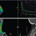
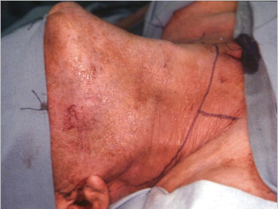
An apron flap incision was made at the level of the thyroid cartilage extending to both sternocleidomastoid muscles ( Fig. 9‑3).
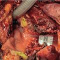
The superior laryngeal nerves were identified and preserved at the midpoint between the superior corner of the hyoid bone and the thyroid cartilage, running along the superior laryngeal artery, a branch of the superior thyroid artery ( Fig. 9‑4).
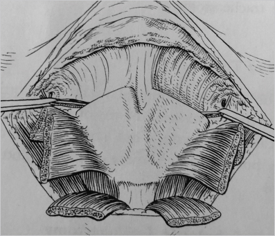
The infrahyoid musculature was skeletonized from the hyoid bone, followed by the transverse section of the thyroid cartilage ( Fig. 9‑5).
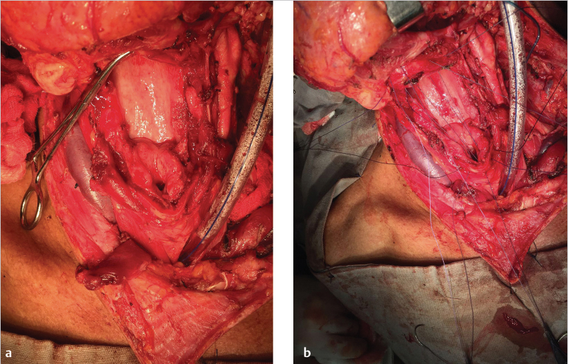
The mucosa was opened at the level of the laryngeal ventricles. A transverse pharyngotomy, and section of the musculature of the base of the tongue were performed. After the pharyngotomy, the cancer was resected with cancer free mucosal margins. Reconstruction was done by the mucosal suture of the pyriform sinus and three separate sites transferring the thyroid cartilage and base of the tongue.
A tracheostomy and a feeding tube were placed at the end of the procedure. The patient was decannulated on the twenty-fifth postoperative day and around same time started to receive an oral diet. Pathological examination revealed squamous cell carcinoma G3: pT2N1M0, R0, LVI0, PN0, and ECS0.
Stay updated, free articles. Join our Telegram channel

Full access? Get Clinical Tree


