12 Locally Intermediate Glottic Cancer: Vertical Partial Laryngectomy
Abstract
Radiotherapy, laser endoscopic excision, and partial laryngectomy are the options in the treatment and of the moderate-stage cancer of the larynx. The decision must consider the patient’s expectations. For the case described here, vertical technique (hemilaryngectomy) and the horizontal technique (horizontal supracricoid laryngectomy) may be recommended. The main advantage of the hemilaryngectomy is the maintenance of the hemilarynx structures not involved by the carcinoma. Probably the cases presenting microscopic infiltration of the thyroid cartilage for cancer would be eligible for transoral laser resection. Vertical laryngectomies are still a good option in cases where endoscopic surgery does not suffice or when it is not available, such as in some developing countries. The need for the reconstruction of the endolarynx by using flaps and grafts allows for a better glottic coaptation. The oncologic outcome is less favorable in cancer extending beyond the confines of the vocal folds, in which supracricoid laryngectomy can be performed.
12.1 Case Report
A 76-year-old Caucasian male, retired stevedore, presented with a 4 month history of progressive dysphonia, followed by episodes of choking on liquids. He stated that he smoked one pack of cigarettes a day for 45 years as well as drinking alcohol socially. He also stated that he was hypertensive and was taking enalapril and atorvastatin for dyslipidemia.
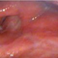
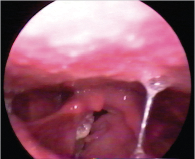
At the time of the consultation, the patient was healthy and presented with hoarseness, with and little vocal range. No abnormalities were found on oroscopy and cervical palpation. A direct laryngoscopy revealed an ulcer-ated infiltrative lesion on the entire right vocal fold, which extended to the anterior commissure, with no involvement of the right vocal fold. The lesion did not extend to the infraglottis, ventricle, or ipsilateral arytenoid ( Fig. 12‑1) The mobility of the left vocal fold was reduced and Reinke’s edema present.
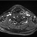
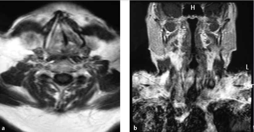
For a better deeper evaluation of the extension of the lesion, an MRI scan was performed which demonstrated an infiltrative irregular lesion of the right vocal fold, with heterogeneous contrast enhancement, with partial invasion of the paraglottic space. The lesion infiltrated the anterior commissure, but the ipsilateral arytenoid was preserved. There were no signs of extension to the left vocal fold ( Fig. 12‑2).
Microlaryngeal surgery for better staging and biopsy was carried out under general anaesthesia. The anatomopathological result showed a moderately differentiated squamous cell carcinoma.
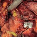
The patient underwent a hemilaryngectomy, with the removal of the wedge of thyroid cartilage in en bloc resection of the cancer, respecting oncological margins in depth in the paraglottic space. Both arytenoids were preserved. Prior to the beginning of the hemilaryngectomy, a protective tracheostomy was performed, at the fifth tracheal ring out of the operative field. For the performance of the hemilaryngectomy, the skin was incised over the thyroid cartilage and the subplatysmal flap was raised. With the opening of the median raphe and exposure of the thyroid cartilage, the fascia and the perichondrium were incised. Thyrotomy was performed vertically, at a distance of 4 mm from the midline contralateral to the cancer and at least 3 mm anterior to the posterior border of the ipsilateral thyroid cartilage. Entry into the larynx was performed by opening the cricothyroid membrane. The thyroid cartilage was retracted laterally and the oncological margins in the mucosa between 2 and 3 mm of the cancer were carefully delimited under direct view. The frozen section report confirmed that the margins were free. The internal perichondrium of the thyroid cartilage wing was removed together with the surgical specimen which included the epiglottic petiole was included in the specimen, since the anterior commissure was found to be invaded by the cancer. The mucosa of the pyriform sinus was mobilized medially to cover the exposed ipsilateral arytenoid ( Fig. 12‑3).

After the cancer resection was completed, the left vocal fold was reconstructed with a bipedicled flap of the ipsilateral sternohyoid muscle (Bailey’s flap), keeping the cranial and caudal pedicles. The flap was 1.5-cm thick and was kept in place by the pedicles, with no need for fixation. A fixation point of the contralateral vocal fold at the epiglottic petiole formed a neocommissure.
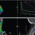
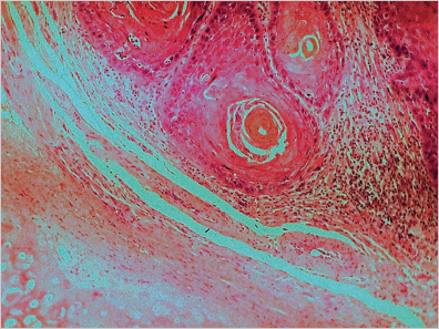
The histopathological report showed well-differentiated squamous cell carcinoma adjacent to the thyroid cartilage, but with no invasion ( Fig. 12‑4).
The patient progressed satisfactorily and the tracheostomy was occluded on the second postoperative day; the cannula was removed 24 hours later, and the patient was discharged. The nasoenteral feeding tube was retained for a total of 5 days, after the reintroduction of purée on the third postoperative day. Patient’s airway was adequate and the voice was rough.
Stay updated, free articles. Join our Telegram channel

Full access? Get Clinical Tree


