Etiology
Prevalence (%)
Laboratory screening
Nonfunctional adenoma
85
Normal
Subclinical Cushing’s syndrome
7
1-mg DST(serum cortisol >3 μ/dl)
Plasma ACTH (<5 pg/ml)
Pheocromocytoma
3.5
24-h urine metanephrines >2 mg/g Cr
Hyperaldosteronism
0.7
Plasma aldosterone ≥15 ng/dl with a APRR ≥20
Nonfunctional adrenocortical carcinoma
2
Normal
Prevalence increases with age, especially in the 5th and 7th decades of life, but is regarded as uncommon in individuals with less than 30 years of age. Hypertension, obesity, and diabetes have been frequently reported, particularly in bilateral lesions, and this may be due to the presence of subclinical hypercortisolism [4].
Imaging Procedures
CT scan is the primary and preferred method used for evaluation of adrenal glands because it is a procedure that is readily available, while offering the highest spatial resolution. Contrast enhancement and deenhancement (washout) in late phases (15 min) are helpful to characterize the lesions; however, noncontrast CT is often sufficient for the diagnosis of AI (Fig. 11.1).
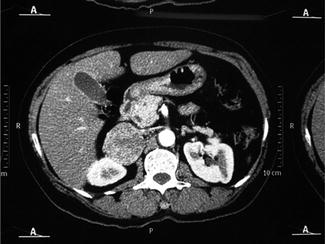
Fig. 11.1
A 52-year-old woman with primary hyperparathyroidism and hypertension had a CT scan to evaluate for renal calculi. A lesion (5.5 cm) in right adrenal gland was discovered. Noncontrast attenuation: 39 HU. Contrast washout: 42 % in 15 min
Among the characteristics for identification of potentially malignant lesions, the first to be observed is the size of the mass. Various cut points have been proposed ranging between 3 and 6 cm (more often 4 cm) of diameter based on the fact that primary carcinomas of the adrenal with measurements lower than these are quite rare. However, adrenal carcinomas of 2.5 cm have been documented, and the use of these cutoff limits can exclude patients harboring carcinomas that are still small, and therefore, offer the greatest likelihood of being able to be treated and cured if operated upon in early stages [5, 6].
The next, and probably the most important, aspect to be seen is related to the noncontrast attenuation of the lesion when evaluated by CT. The rationale is that adrenal adenomas, due to their high content of intracellular lipids, usually exhibit low attenuation. In this regard, using a cutoff level of 10 HU (Hounsfield units) or less, a sensitivity of 96–100 %, and a specificity of 50–100 % have been reported for diagnosing a benign lesion [1, 7]. Several authors reported high sensitivity and specificity in CT after using contrast. Percentage deenhancement, the so-called washout, of at least 60 % in 15 min has been shown to be 98 % sensitive and 92 % specific for benign lesion [7] (Fig. 11.1).
Other features of benign lesions that can be evaluated by CT, such as regular margins and homogeneous attenuation, have low accuracy and are less useful in diagnosis. Calcifications, necrosis, and hemorrhages are atypical events, but occur more specifically in larger lesions.
In adrenal hemorrhage, a clinical condition associated with sepsis, coagulation disorders, adrenal tumors (such as pheochromocytomas), and abdominal trauma, CT scan initially show high density that gradually decreases as the hematoma subsides which may in turn become a pseudocyst. Other lesions with characteristic features include cysts and myelolipomas. Endothelial cysts and pseudocysts are the most common, accounting for more than 80 % of the cases. CT is useful in demonstrating the presence of liquid (hypodense) and generally thick capsules, whereas MRI shows hypointensity on T1, and hyperintensity on T2. Patients with adrenal cysts can, in some cases, benefit from fine needle aspiration (FNA) biopsy guided by ultrasound or CT for decompression and/or cytologic evaluation of the aspirated fluid. Since pseudocysts can originate from pheochromocytomas, measurements of serum or urinary catecholamines or metanephrines are always recommended [3, 5, 6].
Myelolipomas, in turn, are benign tumors composed of mature adipose tissue and normal hematopoietic tissue, corresponding to around 7–15 % of AI cases. They are almost always asymptomatic lesions. Seen by CT, they appear as hypo-dense lesions (−40 HU), due to the presence of fat. Calcifications and hemorrhages may also be present [5]. Bilateral lesions occur in about 20 % of AI cases and are usually due to metastases, granulomatous diseases, adrenal hemorrhage, or congenital adrenal hyperplasia. In this regard, positron emission tomography (PET) using 2-18F-fluoro-2-deoxy-d-glucose, has been used in cancer patients to evaluate the possibility of adrenal metastasis by demonstrating increased uptake, as well as being able to differentiate these cases from adenomas, which may not demonstrate the same uptake [7, 8].
Related posts:

Stay updated, free articles. Join our Telegram channel

Full access? Get Clinical Tree


