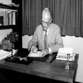
Emil Zuckerkandl. (Circa 1890, author unknown, public domain)
Emil Zuckerkandl was born on September 1, 1849, in Gyӧr, Hungary, and died on May 28, 1910, in Vienna. He was a professor and anatomist whose name is well known to endocrine surgeons for his description of two important anatomical entities: the “tubercle of Zuckerkandl” in the neck and the “organ of Zuckerkandl” in the abdomen.
Zuckerkandl studied medicine at the University of Vienna in 1867, completing his degree in 1874. His younger brother, Otto, also studied medicine and went on to become a professor and urologist in Vienna. Emil was an exceptionally able student who so impressed his anatomy teacher , Joseph Hyrtl, at the Vienna School of Anatomy, that he was appointed as a demonstrator in anatomy, and later sent to the University of Utrecht in Amsterdam as a prosector. In 1873, he returned to Vienna to work with Langer at the Vienna School of Anatomy, the world’s leading anatomy center for over half a century [1]. Following the death of Langer in 1888, he assumed the chair of descriptive and topographic anatomy in Vienna. In 1905, Zuckerkandl edited Heitzmann’s Atlas of Descriptive Anatomy of Man, with over 600 illustrations. As professor, he was noted for his observational powers and critical appraisal. He made significant contributions to normal and pathological anatomy and was known to anatomists and surgeons from around the world as the most indefatigable worker. He published over 160 papers, although the writer of his obituary, published in The Lancet in 1910, did note that he was “a patient and diligent worker, too ready to publish perhaps, and somewhat urgent and short-sighted in his claims for priority” [2].
The organ of Zuckerkandl (Fig. 1) was described by Zuckerkandl in 1901 [3] and comprises a collection of chromaffin cells located between the origin of the inferior mesenteric artery and the aortic bifurcation. It is thought to have a physiologic function in early gestation in relation to blood pressure control; however, it regresses late in the third trimester. Its importance to endocrine surgeons arises from its propensity to be a site in which extra-adrenal pheochromocytoma may develop.
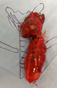
The subsequent year, in 1902, he published his description of the tubercle of Zuckerkandl [4]. Prior to this, he had in fact described aspects of thyroid anatomy during his dissections of the oral cavity , noting in 1878 the presence of “glandular masses with the structure of thyroid, embedded in the tongue above the hyoid” (presumably a lingual thyroid). In his drawings of the tubercle of Zuckerkandl (Fig. 2), a clear appreciation of the relationship between a posterolateral projection of thyroid tissue immediately lying adjacent to the normal position of the superior parathyroid gland can be seen . Although its clinical significance was almost certainly appreciated by Zuckerkandl at the time, knowledge of the structure was lost to endocrine surgeons until “rediscovered” decades later [5]. The tubercle of Zuckerkandl is now considered to be the key to understanding the surgical anatomy of the thyroid and parathyroid glands [6]. The vascular relationship of the tubercle of Zuckerkandl to the superior parathyroid gland and the terminal part of the recurrent laryngeal is critical to safe thyroid lobectomy, [7] while failure to appreciate nodular change within the tubercle of Zuckerkandl (Fig. 3) is a common cause of recurrent disease after total thyroidectomy [8].
Fig. 2
Zuckerkandl’s drawing of the tubercle demonstrating the clear relationship with the superior parathyroid glands. ([4])