Figure 21.1
Orientation of incisions of the lower extremity (a), upper extremity (b), and trunk (c)
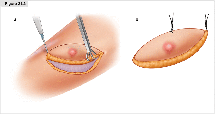
Figure 21.2
(a) Excision of skin and subcutaneous tissue in an ellipse 2 cm from the lesion in the short axis and 6 cm in the long axis. (b) Marking of the specimen with a long stitch on the lateral edge and a short stitch on the superior edge
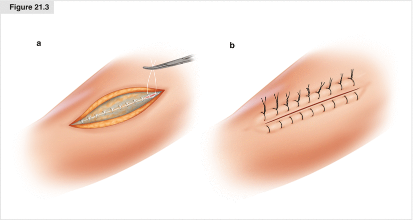
Figure 21.3
(a) Scarpa’s fascia is approximated with interrupted 2-0 Vicryl sutures, then 3-0 nylon vertical mattress sutures are placed but not tied. (b) Closure of the skin after tying nylon sutures
21.2.2 Local Flaps
If cosmesis is a concern, a simple flap may be considered. A rotational flap can be fashioned by drawing a 90° arc from the apex of the defect, then making an incision of several centimeters at a right angle to the base. The tissue is extensively undermined above the level of the muscle fascia, and then rotated into the defect (Fig. 21.4a). Alternatively, a rhomboid flap can be created by making an incision from the edge of the wound for a distance of about three quarters of the width of the wound. Next, an incision of the same length is made from the end of this line at 60°, so that it runs parallel to the wound edge. The tissue between the incisions is freed up from the underlying muscle, keeping the fascia with the flap for improved blood supply. The point at the edge of the wound and the first incision is sutured to the edge of the wound at the opposite side. The point where the two incisions meet is sutured to the apical area of the wound between the first two points, and then the edge of the bottom incision is rotated to the wound edge (Fig. 21.4b). Interrupted nylon sutures are then used to approximate the edges of the rhombus to the circular defect and to close the lateral incision.
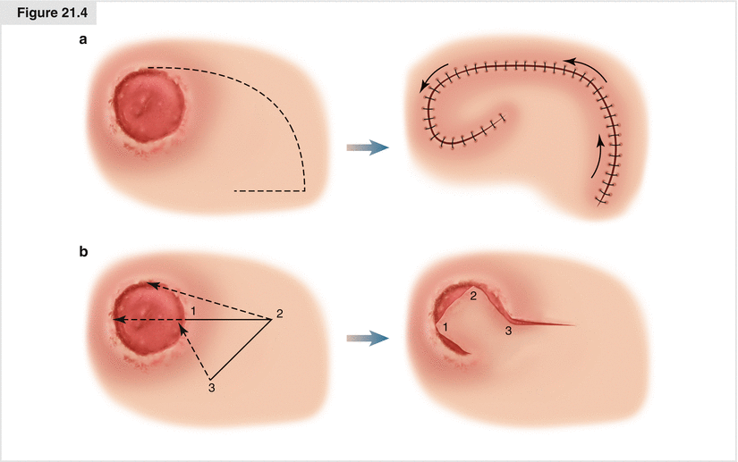
Figure 21.4
(a) Rotational flap: a 90° arc is made laterally and then rotated into place. (b) Rhomboid flap: an incision is made perpendicular to the edge, slightly smaller than the diameter of the wound; the same length incision is made at 60°. Tissue is extensively undermined, then rotated into the defect as shown. The numbers designate the initial and final positions of the corresponding areas of skin.
21.2.3 Sentinel Lymph Node Biopsy
Patients are brought to the Nuclear Medicine suite prior to their procedure, and 0.5–2 mCi (18–74 MBq) of filtered 99Tc-sulfur colloid is injected intradermally in four quadrants around the base of the skin lesion. This injection is generally done within a few hours of surgery, but good results can be obtained even when it is done 12–24 h prior to surgery. Lymphoscintigraphy is performed with a gamma camera to determine the nodal basins draining from the lesion. For extremity lesions, these will be in the corresponding axillary or inguinal areas, only rarely involving the epitrochlear or popliteal regions for distal extremity lesions. For truncal lesions, the drainage is less predictable, and could involve either or both axillae and/or inguinal regions, or even cervical nodes. If two to three nodal basins show uptake on lymphoscintigraphy, then that part of the skin can potentially drain to any of these nodes, so sentinel nodes will need to be dissected out for all of these nodal basins.
The patient is transported to the operating room, where we prefer the use of general anesthesia. Using a 25-gauge needle, 0.5–3 mL of isosulfan blue dye (Lymphazurin; US Surgical-Tyco; Norwalk, CT, USA) is injected intradermally in four quadrants around the primary lesion (Fig. 21.5a). If the needle is truly in the intradermal space, the injection will be very difficult, and a blue wheal will develop slowly at the injection site; if the dye goes into the skin easily, then it is probably being injected subcutaneously, which may lead to less reliable mapping. Patient positioning will then depend on the site of the primary tumor and the draining nodes; we try not to have to reposition patients, but sometimes it is necessary, especially if multiple drainage basins are seen. We generally perform the sentinel node biopsy prior to excision of the primary to reduce the risk of contamination of the instruments, which may occur if the primary is resected first. In some cases, however, especially when the lesion is very close to a nodal basin, the primary must be resected first so that background levels can be reduced and will interfere less with the node counts.
Related posts:


Stay updated, free articles. Join our Telegram channel

Full access? Get Clinical Tree


