Viral Hepatitis


Viral Hepatitis


General Characteristics of Hepatitis


The term hepatitis refers to inflammation of the liver. This chapter discusses infectious hepatitis caused by various viruses.


According to the World Health Organization (WHO), 2 billion people are infected with hepatitis. Almost one third of the world’s population has been infected with one of the known hepatitis viruses. In the United States, acute viral hepatitis most frequently is caused by infection with hepatitis A virus (HAV), hepatitis B virus (HBV), or hepatitis C virus (HCV). These unrelated viruses are transmitted via different routes and have different epidemiologic profiles. Safe and effective vaccines have been available for hepatitis B since 1981 and for hepatitis A since 1995.


Incidence


Primary hepatitis viruses account for approximately 95% of the cases of hepatitis. These viruses are classified as primary hepatitis viruses because they attack primarily the liver and have little direct effect on other organ systems. The secondary viruses involve the liver secondarily in the course of systemic infection of another body system. The viruses for hepatitis types A, B, C, D, E, and GB virus C, as well as secondary viruses (e.g., EBV, CMV), have been isolated and identified (Table 23-1).


Signs and Symptoms


As a clinical disease, hepatitis can occur in acute or chronic forms. The signs and symptoms of hepatitis are extremely variable. It can be mild, transient, and completely asymptomatic or it can be severe, prolonged, and ultimately fatal. Many fatalities are attributed to hepatocellular carcinoma in which hepatitis viruses B and C are the primary causes. The course of viral hepatitis can take one of four forms—acute, fulminant acute, subclinical without jaundice, and chronic (Table 23-2).


Hepatitis A


Etiology


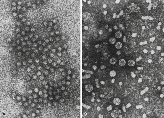
HAV is a small, RNA-containing picornavirus and the only hepatitis virus that has been successfully grown in culture (Fig. 23-1, A). The structure is a simple nonenveloped virus with a nucleocapsid designated as the hepatitis A (HA) antigen (HA Ag). Inside the capsid is a single molecule of single-stranded ribonucleic acid (RNA). The RNA has a positive polarity and proteins are translated directly from the RNA. Replication of HAV appears to be limited to the cytoplasm of the hepatocyte.


The highest titers of HAV are detected in acute-phase stool samples. Human infectivity of saliva and urine from patients with acute hepatitis A does not pose a significant risk. Sexual contact has been suggested as a possible mode of transmission.


Epidemiology


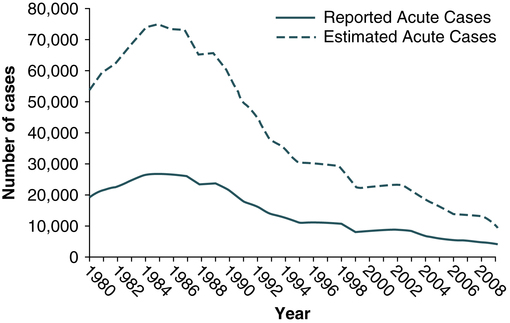
Hepatitis A virus was formerly called infectious hepatitis or short-incubation hepatitis. In developing countries, hepatitis A is primarily a disease of young children; the prevalence of infection, as measured by the presence of antibody (immunoglobulin G [IgG] anti-HAV), approaches 100% at or shortly after 5 years of age. The national rate of hepatitis A has declined steadily since the last peak in 1995 (Fig. 23-2). After asymptomatic infection and underreporting were taken into account, an estimated 21,000 new infections occurred in 2009 (the last year for which statistics were available at the time of publication).


The incidence of hepatitis A varies by age. Historically, the highest rates were observed among children and young adults. Effective vaccines have been available since 1995. Since the issuance in 1999 of recommendations for routine childhood vaccination, rates of hepatitis A have declined. In 2005, the licensing of hepatitis A vaccines was revised to allow vaccination of children aged 12 to 23 months. Nationwide, hepatitis A vaccination of children is likely to result in lower overall rates of infection.


Susceptibility to infection is independent of gender and race. Crowded unsanitary conditions are a definite risk factor. HAV is transmitted almost exclusively by a fecal-oral route during the early phase of acute illness; the virus is shed in feces for up to 4 weeks after infection. Large outbreaks are usually traceable to a common source, such as an infected food handler, contaminated water supply, or consumption of raw shellfish. Institutions and day care centers are known to be favorable sources for transmission as well.


Hepatitis A infection is noted for occurring in isolated outbreaks or as an epidemic, but it also may occur sporadically. Although rarely a transfusion-acquired hepatitis because of its transient nature, an outbreak of HAV infection that occurred in 52 patients with hemophilia in Italy was documented to have been acquired through infusion of contaminated factor VIII concentrate. This concentrate had been treated by a virucidal method (solvent detergent) that ineffectively inactivates nonenveloped viruses.


Improvements in socioeconomic and sanitary conditions and declining family size may be responsible for a decreasing frequency of infection. The incidence of HAV infection is not increasing among health care workers or in dialysis patients. Maternal-neonatal transmission of HAV is not recognized as an epidemiologic entity. Person to person contact, usually among children and young adults, remains the major route of HAV infection.


The most frequently identified risk factor for hepatitis A has been international travel, reported at 15% of patients overall. Most travel-related cases have been associated with travel to Mexico and Central or South America (70%). As HAV transmission in the United States has decreased, cases among travelers to countries where hepatitis is endemic have accounted for an increased proportion of all cases.


Sexual and household contact with another person with hepatitis A have been among the most frequently identified risk factors, reported for 10% of cases in 2006. In 2006, the proportion of HAV-infected persons who reported injection of street drugs was 2.1%.


Signs and Symptoms


Nonimmune adult patients infected with HAV can develop clinical symptoms within 2 to 6 weeks after exposure (average, ≈4 weeks). However, hepatitis A is often a subclinical disease, with many patients being anicteric. Clinically apparent cases show elevated serum liver function enzyme and bilirubin levels, with jaundice developing several days later. Viremia and fecal shedding of virus disappear at the onset of jaundice. Atypical presentations include prolonged intrahepatic cholestasis, relapsing course, and extrahepatic immune complex deposition, all of which resolve spontaneously.


Complete clinical recovery is anticipated in almost all patients. Hepatitis A rarely causes fulminant hepatitis and does not progress to chronic liver disease. Unusual clinical variants of hepatitis A include cholestatic, relapsing, and protracted hepatitis. In cholestatic hepatitis, serum bilirubin levels may be dramatically elevated (>20 mg/dL) and jaundice persists for weeks to months before resolution. In relapsing hepatitis and protracted hepatitis, complete resolution is anticipated.


A chronic carrier state (persistent infection) and chronic hepatitis (chronic liver disease) do not occur as long-term sequelae of hepatitis A. Rarely, injection with HAV may cause fulminant hepatitis, with about 0.1% mortality. Fulminant hepatitis is the most likely complication of coinfection with other hepatitis viruses.


Diagnostic Evaluation


Testing methods for HAV include the following:


The short period of viremia makes detection difficult. Specific IgM antibody usually appears about 4 weeks after infection and may persist for up to 4 months after onset of clinical symptoms. The presence of IgG or total (IgM and IgG) antibody indicates past infection or immunization and associated immunity. The total assay detects IgM and IgG antibodies but does not differentiate between them. The hepatitis A antibody IgM assay is appropriate when acute HAV infection is suspected. Specific IgG antibody apparently protects an individual from symptomatic infection, but specific IgM may increase with reinfection. In the acute phase of HAV, liver function levels (e.g., serum liver enzyme levels) will be elevated and may aid in establishing the diagnosis.


Prevention and Treatment


The first effective control measures to prevent enterically transmitted viral hepatitis resulted from World War II research. In 1945, the following were demonstrated: (1) infectious virus could be transmitted by contaminated drinking water; (2) treatment of the water by filtration and chlorination made it safe to drink; and (3) gamma globulin derived from convalescent-phase serum from patients with hepatitis could protect adults from clinical hepatitis. For 50 years, refining food and water preparation and establishing standards for immune globulin constituted the methods of HAV prevention. An individual who has had close contact with an HAV-infected person should receive passive immunization with immune globulin intramuscularly.


A safe, highly immunogenic, formalin-inactivated, single-dose vaccine is available to prevent HAV infection (Box 23-1). HAV vaccine should be targeted at high-risk groups (e.g., staff in child care centers; food handlers; international travelers, including military personnel; homosexual men; institutionalized patients).


Box 23-1   Hepatitis Vaccine: Questions and Answers


Hepatitis A


Who should receive hepatitis A vaccine?


Some people should be routinely vaccinated with hepatitis A vaccine:


Other people might receive hepatitis A vaccine in special situations:


Hepatitis B


Adapted from Centers for Disease Control and Prevention: Viral hepatitis, 2012 (http://www.cdc.gov/ hepatitis/index.htm).


Universal childhood vaccination may prove to be the most cost-effective method of protecting large populations, both nationally and globally. Routine childhood hepatitis A vaccination is recommended.


In May 2001, the U.S. Food and Drug Administration (FDA) approved a new combination vaccine that protects individuals 18 years of age and older against diseases caused by HAV and HBV. The vaccine, called Twinrix (GlaxoSmithKline Beecham, Philadelphia), combines two already approved vaccines, Havrix (hepatitis A vaccine, inactivated) and Engerix-B (hepatitis B vaccine, recombinant) so that those at high risk for exposure to both viruses can be immunized against both at the same time. Areas with a high rate of both HAV and HBV include Africa, parts of South America, most of the Middle East, and South and Southeast Asia. Clinical trials of Twinrix, given in a three-dose series at ages 0, 1, and 6 months, have shown that the combination vaccine is as safe and effective as the already licensed, separate HAV and HBV vaccines.


Hepatitis B


Etiology


HBV is the classic example of a virus acquired through blood transfusion. It serves as a model when transfusion-transmitted viral infections are considered (see Fig. 23-1, B).


The Australia antigen, now called hepatitis B surface antigen (HBsAg), was discovered in 1966. This discovery, and its subsequent association with HBV, led to the biochemical and epidemiologic characterization of HBV infection.


Hepatitis B is a complex DNA virus that belongs to the family Hepadnaviridae; a member of this family is known as a hepadnavirus. Eight different HBV genotypes with differences in clinical outcomes have been identified. Viral proteins of importance include the following:


The unique structure of the DNA of HBV is one of the distinguishing characteristics of a hepadnavirus. The DNA is circular and double stranded, but one of the strands is incomplete, leaving a single-stranded or gap region that accounts for 10% to 50% of the total length of the molecule. The other DNA strand is nicked (3′ and 5′ ends are not joined). The entire DNA molecule is small and all the genetic information for producing both HBsAg and HBcAg is on the complete strand. During the disease process, viral DNA of HBV is actually incorporated into the host’s DNA.


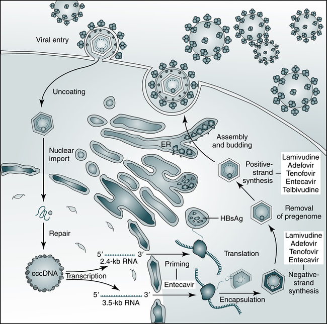
HBV relies on a retroviral replication strategy (reverse transcription from RNA to DNA). Eradication of HBV infection is rendered difficult because stable, long-enduring, covalently closed circular DNA (cccDNA) becomes established in hepatocyte nuclei and HBV DNA become integrated into the host genome (Fig. 23-3).


Epidemiology


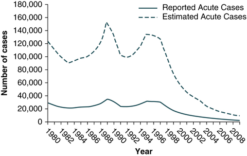
Hepatitis B infection has been referred to as long-incubation hepatitis. In 2009 (the last year for which statistics were available at the time of publication), a total of 3371 acute symptomatic cases and 38,000 estimated total new infections of hepatitis B were reported in the United States. The overall incidence was the lowest ever recorded and represents a decline of 81% since 1990 (Fig. 23-4).


About 1.25 million people in the United States have chronic HBV infection, 20% to 30% of whom acquired the infection in childhood. Each year, about 3000 to 5000 people die from cirrhosis or liver cancer caused by HBV. The highest rate of disease occurs in those ages 20 to 49 years. The greatest decline has occurred in children and adolescents as a result of routine hepatitis B vaccination.


The incidence of HBV infection caused by blood transfusion is increasingly rare in developed countries. Transfusion-acquired HBV has been severely reduced because high-risk donor groups (e.g., paid donors, prison inmates, military recruits) have been eliminated as major sources of donated blood and because specific serologic screening procedures have been instituted. This shift to an all-volunteer donor supply probably accounts for a 50% to 60% reduction of transfusion-related hepatitis. The overall incidence of HBV is high among patients who have received multiple transfusions or blood components prepared from multiple-donor plasma pools, hemodialysis patients, drug addicts, and medical personnel (see Table 23-1).


Persons at risk of exposure to HBV, including those mentioned earlier, include members of the following groups:


Hepatitis B virus does not seem capable of penetrating the skin or mucous membranes; therefore, some break in these barriers is required for disease transmission. Transmission of HBV occurs via percutaneous or permucosal routes and infective blood or body fluids can be introduced at birth, through sexual contact, or by contaminated needles. Infection can also occur in settings of continuous close personal contact. About 50% of patients with acute type B hepatitis have a history of parenteral exposure. Inapparent parenteral exposure involves intimate or sexual contact with an infectious individual. Transmission between siblings and other household contacts readily occurs via transmission from skin lesions such as eczema or impetigo, sharing of potentially blood-contaminated objects such as toothbrushes and razor blades, and occasionally through bites. HBV has been found in saliva, semen, breast milk, tears, sweat, and other biological fluids of HBV carriers. Urine and wound exudate are capable of harboring HBV. Stool is not considered to be infectious.


Signs and Symptoms


Infection with HBV causes a broad spectrum of liver disease, ranging from subclinical infection to acute, self-limited hepatitis and fatal fulminant hepatitis. Exposure to HBV, particularly when it occurs early in life, may also cause an asymptomatic carrier state that can progress to chronic active hepatitis, cirrhosis of the liver, and eventually hepatocellular carcinoma.


A number of factors, including the dose of the agent and an individual’s immunologic host response ability, influence the clinical course of HBV infection. Extrahepatic manifestations, reflecting an immune complex–mediated, serum sickness–like syndrome, are seen in fewer than 10% of patients with acute hepatitis B and include rash, glomerulonephritis, vasculitis, arthritis, and angioneurotic edema. Manifestations such as vasculitis, glomerulonephritis, arthritis, and dermatitis are mediated by circulating immune complex deposition (HBV antigen-antibody) in blood vessels.


The progression of liver disease in HBV infection is fostered by active virus replication, manifested by the presence of an HBV DNA level above a threshold of approximately 1000 to 10,000 IU/mL. Patients with lower levels and normal liver enzyme levels are considered to be inactive carriers, with a low risk of clinical progression. Rarely, reactivation in these patients can occur spontaneously or with immunosuppression. Perinatal infection can result in high HBV level replication without substantial liver injury in the early decades of life; however, the risk of progression to cirrhosis and hepatocellular carcinoma is proportional to the level of HBV DNA maintained persistently over time.


Persistent infection is the usual consequence of HBV infection acquired at an early age, signaled by the prolonged presence of HBsAg. Some individuals with chronic HBV infection are asymptomatic carriers, whereas others have clinical, laboratory, and histologic evidence of chronic hepatitis that may be associated with the development of postnecrotic cirrhosis. Persistent HBV infection is believed to be a precursor of primary hepatocellular carcinoma. In about 5% to 10% of individuals with HBV, especially patients with immunodeficiencies (e.g., AIDS), the disease will progress to a chronic state.


Laboratory Assays


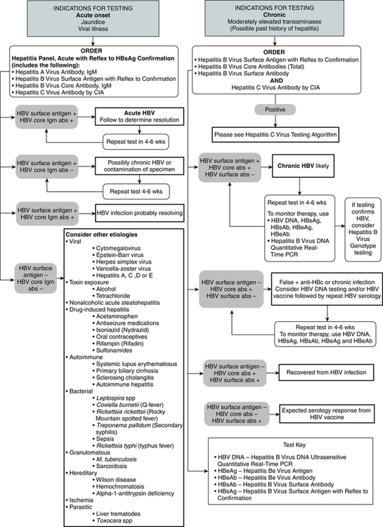
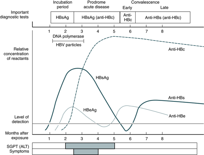
Laboratory diagnosis (Fig. 23-5) and monitoring of acute and chronic HBV infections involve the use of several of the following tests (Tables 23-3 and 23-4):


Serum testing procedures may be performed by qualitative chemiluminescent immunoassay, qualitative EIA, quantitative real-time PCR, quantitative real-time PCR–nucleic acid sequencing, or real-time PCR with reflex to genotype. Immunohistochemistry may be used to detect HBsAg in liver tissue samples.


Hepatitis B Surface Antigen


Serum HBsAg is a marker of HBV infection. Antibodies against HBsAg signify recovery. The initial detectable marker found in serum during the incubation period of HBV infection is HBsAg. HBsAg usually becomes detectable 2 weeks to 2 months before clinical symptoms and as soon as 2 weeks after infection. This marker is usually present for 2 to 3 months. This procedure screens for the presence of the major coat-protein of the virus (HBsAg) in serum and is considered to be the most reliable method of choice for preventing the transmission of HBV via blood. The presence of HBsAg indicates active HBV infection, acute or chronic.


The titer of HBsAg rises and generally peaks at or shortly after the onset of elevated liver serum enzyme levels (e.g., ALT, SGPT). Clinical improvement of the patient’s condition and a decrease in serum enzyme concentrations are paralleled by a fall in the titer of HBsAg, which subsequently disappears. There is variability in the duration of HBsAg positivity and in the relationship between clinical recovery and the disappearance of HBsAg (Fig. 23-6). About 5% of positive HBsAg values are false-positive results.


Among persons infected with HBV with detectable HBsAg in their serum, not all the HBsAg represents complete Dane particles. HBsAg-positive serum also contains two other virus-like structures, which are incomplete spherical and tubular forms consisting entirely of HBsAg and devoid of HBcAg, DNA, or DNA polymerase. The incomplete HBsAg particles can be present in serum in extremely high concentrations and form the bulk of the circulating HBsAg.


Jun 12, 2016 | Posted by in IMMUNOLOGY | Comments Off on Viral Hepatitis

Full access? Get Clinical Tree

Get Clinical Tree app for offline access