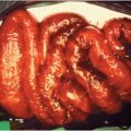
Figure 41.1 Infected atherosclerotic aneurysm of descending thoracic aorta (arrows): blood cultures grew Salmonella. (Courtesy of David Schlossberg, MD.)
Clinical manifestations depend to a large extent on the site of the aneurysm (Table 41.1), although mycotic aneurysms are often clinically unsuspected. Most infected aortic aneurysms occur in elderly atherosclerotic men (4:1 ratio, men > women), but symptoms are nonspecific and may overlap with those of uninfected aneurysms. Fever and continuing bacteremia despite seemingly appropriate antimicrobial therapy are suggestive of an infected intravascular site. CT scan is considered the optimal initial imaging technique, with multiple newer imaging modalities including MRI, FDG-PET, and single-photon emission computed tomography (SPECT) emerging as increasingly feasible and helpful adjunctive imaging techniques.
| Site | Frequency of diagnosis (range) | Clinical presentation | Imaging | Microbiology | Management |
|---|---|---|---|---|---|
| General | |||||
| All infected aneurysms | 100% | Fever common (70%–94%) Malaise, weight loss Pain (100%) Rapidly expanding mass Leukocytosis (65%–85%) Positive blood cultures (50%–75%) | Findings: Aneurysm with lack of intimal calcification Perianeurysmal fluid/gas collection Studies: CT with contrast, MRI Ultrasonography (if accessible) Radionuclide-tagged WBC scans | Staphylococcus 40% (at least 66% S. aureus) Salmonella 20% Streptococcus 20% Escherichia coli 6% IVDU: S. aureus, Pseudomonas spp. Enterococcus spp., Streptococcus viridans | Surgical: Wide debridement, irrigation with antibiotic solution of involved tissues, complete resection of aneurysm if possible Antibiotic: Empiric treatment with IV antibiotics for 6–8 wk after surgery based on culture results of resected tissue Follow-up blood cultures Consider chronic suppressive oral antibiotic therapy when extra-anatomic bypass is not performed (i.e., for in situ repairs) |
| Specifics | |||||
| Aorta Infrarenal abdominal aortaa Ascending aorta and arch (secondary to endocarditis) | 27% (11%–75%) | Abdominal or back pain Palpable abdominal lesions (about 50%–65%) Vertebral osteomyelitis (lumbar/thoracic) | Frontal, lateral abdominal x-ray studies Abdominal ultrasound | Salmonella spp. have predilection for suprarenal aorta Staphylococcus predominates in infrarenal aorta | Extra-anatomic arterial reconstruction (axillofemoral or aortofemoral) If risk too high, in situ reconstruction with cryopreserved allograft |
| Visceral artery Superior mesenteric,a splenic, hepatic, celiac, renal | 24% (0%–29%) | Colicky abdominal pain Jaundice (hepatic artery) Hemoptysis or hemothorax (celiac artery) | Ultrasound may exclude other causes (e.g., pancreatic masses) | Bacteroides fragilis reported from supraceliac aorta and celiac artery | Complete excision may be hazardous; careful drainage and longer-term antibiotic therapy may be necessary |
| Iliac | 4% (0%–25%) | Thigh pain, quadriceps wasting, depressed knee jerk Arterial insufficiency of extremity | Excision and arterial ligation; reconstruction usually can wait until infection has resolved | ||
| Arm Radial arterya Brachial artery Subclavian artery | 10% (0%–9%) | Pain over site of lesion About 90% palpable May appear as cellulitis, abscess; distal embolic lesions; skin changes common | Proximal ligation of the vessel, resection of the aneurysm, and appropriate drainage should be followed by antibiotic therapy. | ||
| Leg Femoral arterya | 12% (4%–44%) | Pain over site of lesion About 90% palpable Pulsatile mass, decreased peripheral pulses Possible local suppuration, distal embolic lesions; petechiae, purpura | S. aureus incidence as high as 65% | Excision and arterial ligation; reconstruction usually can wait until infection has resolved Autogenous grafting may allow reconstruction through the bed of the resected aneurysm if anastomoses performed in clean tissue planes | |
| Intracranial Peripheral middle cerebral arterya | 4% | Usually clinically silent May appear as severe unremitting headache Usually secondary to endocarditis | Four-vessel cerebral arteriography invaluable MRI | Enterococcus spp. S. viridans Pseudomonas spp. Candida albicans |
Abbreviations: CT = computed tomography; MRI = magnetic resonance imaging; WBC = white blood cell; IV = intravenous; IVDU = intravenous drug user.
a Most common site or manifestation.
A variety of intra-arterial prosthetic devices are now being used in cardiovascular medicine, including arterial closure devices, prosthetic carotid patches, coronary artery stents and endovascular stents, and stent-grafts. Infections of these devices remain either uncommon or extremely rare, but infectious complications associated with the placement of these devices are often devastating. S. aureus has been implicated in as many as three-quarters of these cases, and has been the primary pathogen, even in late-onset infections. Blood cultures should be obtained from all patients who have a history of endovascular stent placement and local or systemic signs of infection.
Therapy
Stay updated, free articles. Join our Telegram channel

Full access? Get Clinical Tree


