1
Shared/self-/differentiation TAAs (e.g., Melan-A, PSA, CEA)
2
Shared/self-/cancer testis or germinal TAAs (e.g., MAGE, NY-ESO-1)
3
Universal TAAs (e.g., survivin, hIERI)
4
Mutated, unique TAAs
The TAAs recognized by T cells of the first three subgroups have been characterized (Table 2.1), and their T cell peptide epitope sequence has been defined. Therefore, during the last 10 years or so, such TAAs have lent themselves to clinical use in vaccination trials owing to the possibility to synthesize them in purified form and relevant quantities. This availability was not matched by that of unique, mutated TAAs discovered if not in recent years and used in early animal studies [6, 7]. This circumstance thus led to a discrepancy between self-TAAs and mutated TAAs, since the latter were not available as purified peptides and consequently not usable if not as cellular extracts. Therefore many protocols were carried out with molecularly characterized self-TAAs including the differentiation proteins, the C/T, and the universal ones (see Table 2.1). The advantage of the availability of molecularly defined TAAs, however, was counteracted by their weak immunogenicity since several forms of tolerance to these self-antigens along with tumor immunosuppressive mechanisms (see below) prevented the occurrence of a strong, clinically meaningful reaction [8, 9]. However, an attempt to define a prioritization in the use of TAAs has been made resulting in a useful classification in terms of advantages and disadvantages of the 75 TAAs considered in this publication [10].
2.2 Formulations Used in Cancer Vaccines
Cancer vaccines have been used under several different formulations without unequivocally showing the superiority of one formulation over the others. By taking the past and successful experience gathered from antiviral vaccines, immunological adjuvants were used as local compounds enabling the injected vaccine to recruit inflammatory cells at the site of injection before vaccine degradation by subcutaneous enzymes.
This allows the TAA to persist locally and thus helps antigen-presenting cells to present such antigens to the immune system usually to the lymph nodes driving the region of vaccine injection.
In early trials, cellular vaccines were mostly used in combination with different adjuvants (e.g., BCG, KLH, Montanide) since they were easily obtainable from autologous or allogeneic cancer cells usually after in vitro stabilization of a cancer cell line. This choice was based on the unproved hypothesis that randomly selected neoplastic cells were representative of the antigenic profile of patient’s tumor mass. These cell vaccines usually failed to show therapeutic activity in appropriate phase III trials [11].
Subsequently, cancer cells were genetically modified to express and release gene products (e.g., Chemokines as IL-2, IL-4, IL-7, IL-12, GM-CSF) able to help the in vivo TAA recognition by T cells and their expansion [12]. However, even after such manipulation, no clear and reproducible increase of the clinical response was obtained in phase II studies [12]. The only vaccine that reached the phase III trial in prostate cancer patients (Vital-2 G-Vax, Cell Genesis) was also disappointing since the study had to be discontinued due to more deaths occurring in the vaccinated arm compared to the placebo arm (see Medical News Today, August, 31, 2008). The reason for this imbalance in death was not identified; nonetheless it is known that very high doses of GM-CSF, released by the cell vaccine, may impair rather than increase patients’ antitumor immune response [13]. However, a successful phase III trial was performed during the last few years leading to its approval by the FDA (Provenge) (see below).
2.2.1 Peptide-/DC-Based Vaccination Against Cancer
As soon as TAA peptides recognized by T cells became available and taking into consideration the ineffectiveness of different B cell-defined TAA to generate widespread tumor cytotoxic antibody response, several phase I–II clinical studies were initiated to assess safety and immune and clinical response in cancer patients, particularly in melanoma-bearing subjects since this neoplasm is considered to be immunogenic [14]. The formulation of these peptide-based cancer vaccines was, however, quite different going from peptide admixed with immunological adjuvants like Freund’s incomplete adjuvant-like vaccines (e.g., Montanide) to peptide loaded onto autologous dendritic cells. Moreover, short peptides (8–10 aa) were the first to be used in the clinic, whereas long peptides (13–18aa) were used later on since long peptides were described to be more immunogenic as compared to short ones even within the HLA class I restriction [15].
No major safety problem was found in vaccination protocols based on the use of one or multiple peptides selected in vitro for their ability to interact with MHC-specific molecules forming a molecular complex recognizable by patient’s T lymphocytes through their TCR. In fact, peptides deriving from C/T or differentiation TAAs (e.g., MAGE-1/3, MART-1, gp100, CEA, PSA) were widely used in different human tumors, and while TAAs-specific T cell response could be generated in 20–80 % of subjects, the clinical outcome remained limited in terms of tumor response (5–20 %) revealing that T cell response induced by the vaccine was not sufficiently strong and/or durable to generate an effective clinical response in the majority of patients [16]. A summary of these results is provided in Table 2.2 which deals with metastatic melanoma; however, it is also representative of other human neoplasms (e.g., colorectal cancer, NSCLC, H/N, prostate cancer, and glioblastoma).
Table 2.2
Results of first generation (1998–2006) of self-peptide-based vaccination of metastatic melanoma patients (phase I–II studies)
Type of peptide TAA | No. of patients | Clinical response (CR+PR) (mean %) | Immune response (%) |
|---|---|---|---|
Lineage related (e.g., Melan-A) | 159 | 14 | 20–65 |
Cancer/testis (e.g., MAGE) | 92 | 17 | 30–50 |
DC peptides | 124 | 16 | 56 |
DC lysates | 106 | 18 | 46 |
2.3 Factors That May Impair the Immune Response Against Tumors
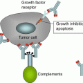
A major problem that came out from the many early studies was the lack or weakness of immune response in mice and patients receiving self-TAAs as vaccines usually combined with immunological, nonspecific adjuvants (e.g., Montanide). A large number of studies were therefore conducted in the last two decades pioneered by the observation of Ferrone’s and Garrido’s groups [17, 18] on the downregulation of class I and II HLA on most tumor cells of a variety of human solid tumors like melanoma, colorectal cancer, etc. This alteration will remove crucial molecules necessary for the TAA presentation to the patient’s immune system. Table 2.3 lists the many different mechanisms of tumor escape that have been described in the last few years and that could have prevented an efficient immune response specifically targeting the appropriate TAAs expressed by cancer cells [19]. A new possibility has been reported in melanoma by different authors describing that BRAF-MAPK signaling is generating cancer-immune evasion [20, 21] though the reverse (i.e., immunostimulation) has been more recently found [22, 23]. In the last few years, the main focus was on the role of myeloid-derived suppressor cells (MDSC) [24, 25] and Tregs [26] as main mechanisms of immune escape by tumor cells. A recent adjunct to the plethora of escape mechanisms is the inflammasome component Nirp3 underlining the complex relationship between inflammation and cancer [27] that impairs vaccine-induced immunity. However, one should consider that several mechanisms may be activated by tumor cells either simultaneously or one after the other according to modifications that occur in the tumor microenvironment and in different tissues in which tumor cells are growing even during different types of therapy [28]. A recent additional mechanism of tumor escape has been described that includes tumor cell secretion of sterol metabolites (LXR ligands), which inhibit the expression of CCR7 on the cell surface of dendritic cells (DCs), thereby disrupting DC migration to the lymph nodes and dampening the antitumor immune priming event [29]. Presently, the major research efforts are directed to find compounds that may help in restoring the full anticancer potential of the immune system [19, 30].
Table 2.3
Factors that interfere with the T cell-mediated antitumor response
Tumor (immunosubversion) | Immune system |
|---|---|
Lack of or downregulation of HLA | Immune anergy or ignorance |
Dysfunction of antigen presentation | Lack of tissue homing molecules |
Release of immunosuppressive factors (IL-10, TGF-beta, VEGF) | T cell receptor dysfunction |
Tumor counterattack (Fas/FasL) | Inactivation of T cells within the tumor environment (granzyme B) |
IDO, SPARC, Galectin3 | Expression of FoxP3, CTLA4, Treg cells |
Endoplasmic reticulum stress | MDSC |
NFAT1, exosomes | Epithelial/mesenchymal transition |
Acidic microenvironment | Tie+ monocytes dysfunction |
2.4 New More Successful Clinical Studies of Vaccination in Cancer Patients
During the last few years, however, new knowledge has been gained on the mechanisms of antitumor immunization and tumor immune escape in cancer patients enrolled in earlier vaccination protocols. Such new information has allowed to overcome some of the hurdles that were identified earlier. Thus several phase II and even phase III vaccination protocols came to their conclusion or to an advanced ad interim evaluation stage and provided clinical evidence of benefit for patients with melanoma and other tumors (Table 2.4). Of note, a phase III protocol of vaccination for metastatic prostate cancer patients with autologous dendritic cells loaded with a hybrid molecule made of GM-CSF and PAP (Provenge) [31] was approved by the FDA.
Table 2.4
Evidence for clinical activity of cancer vaccines
Vaccine
Related posts:Stay updated, free articles. Join our Telegram channel
Full access? Get Clinical Tree
 Get Clinical Tree app for offline access

|
|---|