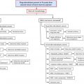
Treatment of iron overload requires robust estimates of total-body iron burden and its response to iron chelation therapy. Compliance with chelation therapy varies considerably among patients, and individual reporting is notoriously unreliable. Even with perfect compliance, intersubject variability in chelator effectiveness is extremely high, necessitating reliable iron estimates to guide dose titration. In addition, each chelator has a unique profile with respect to clearing iron stores from different organs. This article presents the tools available to clinicians to monitor their patients, focusing on noninvasive magnetic resonance imaging methods because they have become the de facto standard of care.
Key points
- •
Serum ferritin and transferrin saturation remain valuable in tracking the therapeutic response to iron-removal therapies.
- •
These inexpensive techniques have many shortcomings that preclude using them safely as sole monitors for chelator efficacy.
- •
Magnetic resonance imaging has become the de facto gold standard for tracking iron levels in the body because it is accurate, reproducible, and well tolerated by patients, and can track iron levels in different organs of the body.
- •
The latter characteristic is important because the mechanisms and kinetics of iron uptake and clearance vary across somatic organs.
- •
The author’s clinical practice is presented as a reference, but individual experiences will still be colored by local expertise as the technologies continue to mature and be more widely distributed.
Related posts:


Stay updated, free articles. Join our Telegram channel

Full access? Get Clinical Tree


