Median sensitivity (range) (%)
Median specificity (range) (%)
Hypoechoic vs. surrounding parenchyma
81 % (48–90 %)
53 % (36–92 %)
Hypoechoic vs. strap muscles
41 % (27–59 %)
94 % (92–94 %)
Absence of Halo
44 % (26–53 %)
89 % (69–98 %)
Microcalcifications
10 % (2–17 %)
94 % (84–98 %)
Macrocalcifications
66 % (33–100 %)
43 % (30–77 %)
Irregular, microlobulated margins
55 % (17–84 %)
80 % (62–85 %)
Solid consistency
86 % (78–91 %)
48 % (30–58 %)
Taller than wide shape on transverse view
48 % (33–84 %)
92 % (82–93 %)
1.1.3.1 Echogenicity
The echogenicity is the capacity to produce echoes and is proportional to the interfaces present in tissue, more interfaces more echogenicity. A rich cell structure contains less interfaces than a tissue with various components (colloid, cells, etc.) and this is the reason because the more aggressive malignant histotype (solid) appear more hypoechoic than those, more differentiated histotype, in which are present the follicular structures. The determination of echogenicity is usually carried out by comparison with the adjacent parenchyma but in some situations this comparison is rather difficult: in Hashimoto’s thyroiditis parenchyma is diffusely hypoechoic. New softwares are developing to quantify natural echogenicity.
1.1.3.2 Calcifications
There are different categories of calcifications:
1.
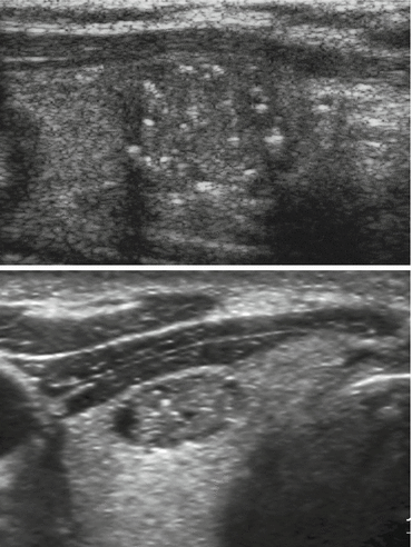
Microcalcifications are small echogenic foci without posterior acoustic shadowing and it is hypothesized that are the ecographic equivalent of psammoma bodies (laminate calcific spherule) that are typical of papillary cancer but occasionally found in Hashimoto diseases or benign nodules. Often in ultrasound reports are described microcalcifications within nodules but in fact these small hyperechoic foci are related to the presence of dense colloid within microcysts associated with the reinforcement of the distal wall (Fig. 1.1).


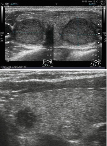
Fig. 1.1
Benign and malignant nodule. (Top) Benign nodule: definite margins with thin and regular hypoechogenic halo (Bottom) Malignant nodule: irregular and ill-defined margins without peripheral halo
2.
Macrocalcifications are larger than 2 mm and causes posterior acoustic shadowing. They may occur either in benign or malignant lesions within areas of fibrosis, tissue degeneration or necrosis.
3.
Peripheral calcifications (“egg shell”) indicate benign lesions in majority of cases but the interruption of peripheral rim may be a sign of cancer invasion [10].
1.1.3.3 Margins
The new technologies for the transducers (high frequency broad band probes) allow a superb definition of margins of thyroid nodules. The presence of ill-defined margins or microlobulated borders is the sign of infiltrating neoplasia (Fig. 1.2). However, may be present indistinct margins in small hyperplastic nodules that should be not confused for infiltrative.


Fig. 1.2
(Top) True microcalcifications. (Bottom) Linear hyperechogenic spots due to dense colloid content in microcysts simulating microcalcifications
1.1.3.4 Halo
Halo is hypo-anechoic rim that surrounds a nodule and is commonly interpreted as the image equivalent of compression of perinodular vessels e/o peripheral edema. Hyperplastic nodules grow very slowly and so displace the surrounding thyroid parenchyma bundling blood vessels in the peripheral portions, as demonstrated by color Doppler. Thin peripheral halo is present in about half of benign lesions but is less common in malignant nodules. Thick irregular halo corresponds to the capsule in benign neoplastic nodules.
1.1.3.5 Vascularization
Initially the amount of intralesional vascularity was associated with malignant nodules but the most recent studies have demonstrated, in small papillary cancer, poor intralesional vascularization and a possible explanation of this discrepancy should be related to the specific pattern of neoangiogenesis in this histological type (anarchical texture, tortuous vasa and A/V shunts). Instead the follicular cancer may be more frequently associated with evident intralesional vascularity. The best method to study the internal vascularization of thyroid nodules is the broadband color Doppler because the “normal” color Doppler tends to overwrite the vascular signal giving the impression of a greater intralesional vascularity than the real one; alternatively you can use the power Doppler. Currently are available volumetric probe that allow the real time tridimensional study of parenchymal vascularization: this method demonstrates the real vascular network inside the thyroid nodule avoiding treated as internal vessels those taken enfilade at the outer edge of the nodule.
1.1.3.6 Taller Than Wide
The ratio between anteroposterior and transverse diameter in thyroid nodules (measured on axial scan) may be indicator of malignancy as in the sonographic evaluation of breast carcinoma. The disproportionate growth in the anteroposterior dimension may be interpreted as an aggressive growth pattern across rather than within normal tissue force lines. The benign nodules that can mimic this pattern are respectively meta-hemorrhagic cyst and outcomes of focal thyroiditis.
1.1.3.7 Stiffness
The malignancy is often associated with stiffness. The US elastography is a novel method that evaluates the tissue elasticity through analysis of radiofrequency shift during manual compression. The stiffness alone is not an indicator of malignancy but is an additional tool along with other ultrasound parameters. Ueno has codified a subjective scale with arbitrary color to differentiate benign from malignant lesions: different degrees of tissue elasticity can be color-coded in parametric images making suspicious tissue changes visible in the ultrasonic image. Recently are disposable semiquantitative and quantitative methods to classify the stiffness of a thyroid nodule. The most promising tool is acoustic radiation force impulse (ARFI) that analyzes the velocity of transmission of shear waves through a discrete lesion determining his stiffness in kilopascal (kP).
1.1.3.8 Associated Features
Evidence of pathological lymph node at cervical level and interruption of thyroid capsular profile are ultrasound features that, if associated with the presence of thyroid nodules, may be diagnostic for malignancy.
Stay updated, free articles. Join our Telegram channel

Full access? Get Clinical Tree


