35 Tumors of the Lung, Pleura, and Mediastinum
Epidemiology and Etiology
Lung cancer is the leading cause of cancer mortality for men and women, exceeding the mortality of breast, colon, and prostate cancer combined.1 In the United States, more than 219,000 new cases of lung cancer and 159,000 deaths were anticipated in 2009.1
The epidemiologic data on smoking and lung cancer fulfill the criteria for causal association, including the consistency of results across studies, the strength of relationship, its specificity, the correct temporal sequence between exposure and disease, and the coherence of the association as evidenced by a dose-response relationship.2 It is estimated that cigarette smoking is the cause of 90% of all lung cancers.3 Multiple genetic alterations of tumor suppressor genes and oncogenes can lead to the pathogenesis of lung cancer. Carcinogens in cigarette smoke are involved in these genetic alterations, mainly through the formation of deoxyribonucleic acid (DNA) adducts.4 There is a clear dose-response relationship between the risk of lung cancer and the number of cigarettes smoked per day, the degree of inhalation, and the age at initiation of smoking.5 The relative risk of developing lung cancer decreases as the time from cessation increases.5
Approximately 10% of lung cancers are thought to be caused by carcinogens other than cigarette smoking. Among nonsmokers, 30% of lung cancer deaths are attributed to radon exposure.3 Increases in lung cancer risk also accompany exposure to carcinogens such as asbestos, bis(chloromethyl) ether, polycyclic aromatic hydrocarbons, chromium, nickel and inorganic arsenic compounds.6,7 The association with occupational exposure to these agents appears to be independent of cigarette smoking. In addition, a number of occupations with high smoking prevalence have increased cancer risk. Women who work as waitresses, cashiers, nurses’ aides, and orderlies have smoking prevalences in excess of 40%, as do men who work as drivers, construction workers, painters, mechanics, and watchmen.8
Chemoprevention strategies have failed to demonstrate the ability to reduce the risk of developing lung cancer. Epidemiologic and case control trials previously reported an inverse relationship between the incidence of lung cancer and the intake of many food groups, including dietary carotenoids such as beta-carotene and lycopene.9 The mechanism of this interaction was theorized to be related to scavenging of free radicals or through antioxidative effects. However, the Beta-Carotene and Retinol Efficacy Trial (CARET), which included more than 18,000 participants, found that supplementation with betacarotene actually increased the risk of developing lung cancer by 12% versus patients who received placebo, and even increased all-cause mortality by 8%.10 Likewise, the retinoid isotretinoin was ineffective in reducing the development of second primary tumors in 1166 patients with prior stage I non–small cell lung cancer (NSCLC), and even increased the risk of death for active smokers.11
Pathogenesis and Histologic Findings
Pathogenesis
There is increasing evidence that lung cancer is derived from a pluripotent stem cell that is capable of expressing a variety of phenotypes. This epithelial stem cell, in normal histogenesis, differentiates into those cells found in the tracheobronchial tree, including pseudostratified reserved cells, ciliated goblet columnar cells, and neuroendocrine cells as type I and II pneumocytes seen lining the alveoli. Those cells that are capable of division can express hyperplastic, metaplastic, or neoplastic change. Lung cancer may exhibit two or more histologic patterns; the frequency of this occurrence may depend of the number of sections examined. In one study, when at least 10 blocks from each tumor were examined, 45 of the 100 cases showed elements of both squamous cell carcinoma and adenocarcinoma.12
Clinical Presentation
Routine screening for lung cancer is being investigated once again. Prior trials conducted in the 1970s failed to show any benefit for the use of chest x-ray examination to screen smokers for lung cancer. More recent studies, however, are exploring the use of computed tomography (CT) scans for screening because they can detect smaller lung nodules. The Early Lung Cancer Action Project was a pilot study of 1000 patients older than the age of 60 with greater than 10 pack-years of smoking who underwent routine screening with low-dose thoracic CT scans. It found almost four times as many cancers as chest x-ray examination alone and these lesions were six times as likely to be early stage and therefore more curable.13 A follow-up study, the International Early Lung Cancer Action Project screened 31,567 asymptomatic persons at risk for lung cancer from 1993 through 2005.14 Of these, 27,456 had repeat screenings 7 to 18 months later. Of the 31,567 participants, 484 were diagnosed with lung cancer, 85% of whom were clinical stage I. The estimated survival in this subgroup was 88%. Other groups have concluded that screening increases the rate of lung cancer diagnosis and treatment, but doesn’t reduce the risk of advanced lung cancer or death from lung cancer.15 Currently, a large national trial is underway to rigorously assess the benefit of screening in this population.
Diagnostic and Staging Procedures
Imaging Studies
Chest x-ray examination remains an extremely valuable tool in the diagnosis of lung cancer. However, CT scan has enormous advantages in its ability to detect lesions that cannot be visualized on chest x-ray examination. It also plays an important role in staging lung cancer, especially spread to areas of the mediastinum undetected on plain films. Typically, a lymph node is considered normal if it is less than 1 cm in diameter on its short axis. CT may also suggest possible areas of local invasion of the primary tumor to the chest wall, vertebrae, or mediastinal structures. Small pleural effusions or pleural nodules, often undetected on plain films, may be evident on CT. A CT of the chest for lung cancer evaluation should also include part of the upper abdomen, which allows for evaluation of the liver and adrenals for metastatic disease. Scanning with 18-fluorodeoxyglucose positron emission tomography (FDG-PET) scanning can also detect neoplastic activity. Studies have shown CT and PET to be more specific and sensitive than PET alone.16 Generally, the sensitivity, specificity, and accuracy of CT scan alone is approximately 70%. CT and PET together have an accuracy of approximately 90% in single-institution studies, although these results may worsen when the technique is studied across multiple institutions. Magnetic resonance imaging (MRI) has not been shown to be more effective than chest CT for lung tumor, but may be helpful to evaluate invasion of vertebral bodies in the mediastinum.
Bronchoscopy
Endobronchial ultrasonography allows detailed imaging of the bronchial wall and peribronchial mediastinum, and may also add to the evaluation of endobronchial lesions.17
Endobronchial ultrasound-guided biopsy of mediastinal and hilar lymph nodes is becoming more commonly employed in the staging of lung cancer. Yields of this technique are approximately 95% with minimal to no severe toxicity.18
Mediastinoscopy and Mediastinotomy
Mediastinoscopy is the most accurate technique to assess superior mediastinal lymph nodes, which are frequently involved in NSCLC. The procedure is simple, safe, and effective in experienced hands. In one large series, the complication rate from cervical mediastinoscopy was 2.3% and the mortality was 0%.19 In patients suspected of having inoperable disease by virtue of mediastinal involvement as detected by CT scanning, confirmation by mediastinoscopy is indicated. For patients who are potentially operable, it is especially important to sample contralateral mediastinal lymph nodes to determine if N3 disease is present. Involvement of aorticopulmonary lymph nodes are typically assessed by anterior mediastinotomy (Chamberlain procedure).20 It is important to remember that these are first-echelon mediastinal lymph nodes for left lung tumors, especially from the left upper lobe, and they may not be sampled when a cervical mediastinoscopy is performed.
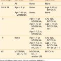
Staging of Lung Cancer
In 1997, the sixth edition of the tumor-node-metastasis (TNM) staging system for NSCLC was published.21 Some criticisms of the staging included that the sample size was relatively small, the data was from few institutions, the staging was mostly surgical, modern imaging was not incorporated into the staging, and there was minimal external validation. In an attempt to improve on these shortcomings, the International Association for the Study of Lung Cancer (IASLC) revised the TNM staging system in 2009.


Stay updated, free articles. Join our Telegram channel

Full access? Get Clinical Tree


