Fig. 20.1
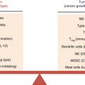
Two mechanisms proposed to explain the association between TAMs and tumorigenesis. (a) A large set of chemokines (CCL2 and others) and cytokines (G-CSF and so on) secreted by tumor cells can promote the recruitment of monocytes in local region and then educate these filtrated monocytes to become TAMs in the location. (b) The inflammatory cytokines produced by TAMs can influence the proliferation of tumor cells. When the factors produced by M2-like TAMs are preponderated, tumor proliferation increases, while the factors produced by M1-like TAMs (reeducated TAMs) are inhibitory for tumor proliferation
20.3 Development of Myeloid Lineage Cells Including Macrophages
Tissue macrophages are divided into two types; nonetheless, some overlap exists in surface marker expression between these two types of macrophage [27]. M1 macrophages (classically activated macrophages or inflammatory macrophages) act essentially to defend the host from a variety of bacteria, protozoa, and viruses and have roles in antitumor immunity. On the other hand, M2 macrophages (alternatively activated macrophages) exert anti-inflammatory properties and can promote wound healing [28]. From the view of functional features, TAMs are overtly similar to M2 macrophages. Tissue macrophages in adults are usually believed to be recruited from monocytes in blood vessels, while monocytes are derived from hematopoietic stem cells (HSCs) in bone marrow (BM). Two types of monocytes have been classified. LY6C hi monocytes (inflammatory monocytes) expressing CCR2 are recruited to acute inflammatory tissues and become M1 macrophages there [29], whereas LY6Clow monocytes (patrolling monocytes) expressing CX3Cl1 are recruited to and become M2 macrophages in tissues usually with chronic inflammation [30]. Recently, the previously believed notion that the origin of adult macrophages are stemmed from HSCs in BM has been challenged, since it is reported that macrophages impositioned vested in the yolk sac (YS) from day 8 (E8) in murine embryo [31], whereas definitive HSCs appeared in the hematogenic endothelium of the aorta-gonado-mesonephros region at E10.5 [32–34] and then migrated to the fetal liver [35]. As shown by Schulz et al., YS-derived F4/80 bright macrophages repopulate in adult tissues and turn to liver Kupffer cells, epidermal Langerhans cells, and brain microglia-independent HSCs [36]. Why do macrophages exist during fetal development in limited organs but in almost all adult tissues is an open question. A possible pathway through which macrophages play their role in development is through guiding morphogenesis [37]. A well-studied example is the mammary gland. Mammalian mammary ducts develop multilaminate bulbous termini known as terminal end buds (TEBs) at puberty and during pregnancy. Macrophages are found within the TEB structure, where they phagocytose apoptotic epithelial cells alone with lumen formation [38, 39]. TAMs may have similar properties but play a role in tumor development instead of tissue development. The vertebrate immune system has evolved in concert with parasites, protozoa, bacteria, and virus infection. A situation faced today is that although the parasite infection has decreased largely for human beings, our immune system against parasites still works actively for allergy reaction, wound healing, and others. Herein, the recent discovery about helminth immunity is briefly narrated. Several kinds of cells participating in helminth immunity should be mentioned ahead; the first cell type which must be pointed is T helper 2 (Th2) cells secreting IL4 in gut or lung when helminth infection occurs. The second kind of cells is gut epithelial Goblet cells, which express IL4Ra, secretory mucus and produces resistin-like molecule-β (RELMβ), an innate protein with direct anti-helminth activity. The third one is M2 macrophages, which own IL4Ra and produce arginase 1, chitinase 3-like proteins 3 and 4 (also known as YM1 and YM2, respectively), and RELMα. Since high arginase activity of myeloid cells coincides with the transport of extracellular L-arginine into cells, causing a reduction of L-arginine in the microenvironment, this decrease in L-arginine would result in T cell hyporesponsiveness [40]. The same thing happens in TAMs. For example, as reported by Rodriguez et al., a subpopulation of mature tumor-associated myeloid cells express high levels of arginase I in 3LL murine lung carcinoma model, and L-Arg depletion by tumor-associated myeloid cells inhibited antigen-specific proliferation of T cells [41]. Despite the high activity of arginase-induced L-Arg depletion, macrophages can convert L-Arg to inducible nitric oxide synthase (iNOS) by other mechanism, which will be discussed later.
Bacterial infection induces macrophage activation, which first recruit neutrophils to the infected site. Neutrophils and macrophages phagocyte the bacteria inside the phagolysosome and kill the bacteria by enzymes inside the lysosome or by reactive oxygen species (ROS) and then produced nitric oxide (NO) radicals. T lymphocytes in regional lymph nodes are stimulated by dendritic cells, followed by the clonal expansion and the migration of these T lymphocytes to infected sites. Among these T cells, Th1 cells produce IFNγ to kill the bacteria inside the phagocytes; Th17 cells produce IL-17 to recruit more neutrophils to the infected site. However, excessive or continued activities of phagocytes and T cells may induce tissue damages and fibrosis, thereby suppressing tissue regeneration. Early studies showed that macrophages can suppress T cell proliferation by producing NO radicals [42, 43] and indoleamine 2, 3-dioxygenase (IDO) [44]. This T cell suppressive function of macrophages is one of TAM characteristics. These macrophages in tumor are specifically called myeloid-derived suppressor cells (MDSCs) [45]. Recently, M2 macrophages have been divided into M2a, M2b, and M2c subgroups according to their inducing stimuli. M2a (induced by exposure to IL-4 and IL-13) and M2b (induced by combined exposure to immune complexes and TLR or IL-1R agonists) exert immunoregulatory functions and drive type II responses, whereas M2c macrophages (induced by IL-10) are more related to the suppression of immune responses and tissue remodeling [46].
20.4 Characteristics of TAMs
Tumor-associated macrophages have been shown to perform a number of different roles in the tumor microenvironment to facilitate tumor progression [37, 47–49], and the density of TAMs in human tumors closely correlates with poor prognosis [5]. TAMs are recruited as monocytes from the bloodstream into tumor tissue. Some chemoattractants produced by both malignant cells and stromal tumor compartments play an important role in this recruitment [50, 51]. For example, stromal- and epithelial cell-produced CSF1 seems the most important chemoattractant working for the recruitment of TAMs to tumor [52], while Csf1 deficiency in macrophages suppressed tumor progression in the mice intestinal cancer model with APC716 mutation [53]. Up to now, various features of TAMs have been identified; however, other features remain to be elucidated. One of these is the close relationship of TAMs and tumor angiogenesis, since TAMs express various angiogenic molecules, including VEGF [54]. Macrophages also promote intestinal cancer by producing TNF, which activates Wnt-catenin pathway essential for tumor progression in intestinal cells [53]. Moreover, TAMs downregulate the expression of major histocompatibility complex class II (MHC II) and their ability of antigen presentation. As for cytokine production, TAMs express COX2-derived prostaglandin E2, as well as the anti-inflammatory cytokine IL-10 [55]. Murine TAMs express low levels of IL-12 but high levels of M2-specific genes, such as arginase-1 (Arg-1), macrophage galactose-type C-type lectin–2 (Mgl2), Fizz1, and Ym1 [56, 57]. These characteristics are similar to M2 macrophages. However, TAMs express both M1 and M2 markers in certain circumstances, relevant to tumor type and the stage of tumor development. For example, increased expression of inducible nitric oxide (iNOS or NOS2, an enzyme expressed by M1 macrophages) together with elevated levels of Arg-1 (usually expressed by M2 macrophages) were observed in TAMs in CT26 murine colon tumors, Meth A− sarcoma, and prostate tumors [58, 59]. Meanwhile, TAMs are thought to suppress T cell proliferation or induce regulatory T cells by the expression of IL-10, TGFβ, Arg-1, and prostaglandins [60–63]. These immunosuppressive macrophages are called myeloid-derived suppressor cells (MDSCs). MDSCs are increased in patients with head and neck, breast, non-small-cell lung, and renal cancers [64–66]. Phenotype of murine MDSCs is CD11b+, Gr-1+, IL-4α+, F4/80−.
20.5 “Reeducating” TAMs to Cytotoxic Phenotype
Due to the large population of TAMs existing in many tumors, a therapeutic approach increasing their tumoricidal activity and attempting to activate antitumor immunity would be most appealing. As previously mentioned, NF-κB signaling pathway is important for cancer-related inflammation and malignant progression. Hagemann et al. stated that the infection of TAMs with Adv-IKKβDN to isolated CD11b+ TAMs from ID8 ovarian cancer-baring mice inhibited NFkβ signaling, and the inactivation of IKKβ in TAMs also prevented tumor cell invasion through macrophage-mediated tumoricidal activity in vitro. Moreover, they demonstrated that IL-12high IL-10low phenotype of IKKβ-targeted macrophages was associated with decreased expression of arginase-1 and elevated expression of inducible nitric oxide synthase (NOS2). They also showed that adoptive transfer of converted tumor by Adv-IKKβDN in vivo induced IL-12-mediated increase in NK cells [67]. Another line of evidence revealed that inhibition of COX-2 can prevent breast cancer metastasis. This was recognized based on the fact that the specific inhibitor of COX-2, etodolac, inhibited human M2 macrophage differentiation, as evidenced by the decreased expressions of CD14 and CD163 genes and increased TNFα production. Using a BALB/c breast cancer model, Na et al. found that etodolac significantly reduced lung cancer metastasis, possibly due to the increased expressions of IA/IE and TNFα genes and decreased expressions of M2 macrophage-related genes [68].
20.6 Concluding Remarks
TAMs have been shown to enhance tumor invasion, migration, and angiogenesis by inflammation. Recent progresses to elucidate the molecular mechanisms of the functions of TAMs opened the new ways to treat cancer patients by reeducating TAMs to be tumor inhibitory cells.
References
1.
2.
3.
Buechler C, Ritter M, Orso E, Langmann T, Klucken J, Schmitz G. Regulation of scavenger receptor CD163 expression in human monocytes and macrophages by pro- and anti-inflammatory stimuli. J Leukoc Biol. 2000;67(1):97–103.PubMed
4.
5.
6.
Leek RD, Lewis CE, Whitehouse R, Greenall M, Clarke J, Harris AL. Association of macrophage infiltration with angiogenesis and prognosis in invasive breast carcinoma. Cancer Res. 1996;56(20):4625–9.PubMed
7.
Lee AH, Happerfield LC, Bobrow LG, Millis RR. Angiogenesis and inflammation in invasive carcinoma of the breast. J Clin Pathol. 1997;50(8):669–73.PubMedCentralPubMedCrossRef
8.
9.
10.
Sfanos KS, De Marzo AM. Prostate cancer and inflammation: the evidence. Histopathology. 2012;60(1):199–215.PubMedCentralPubMedCrossRef
Related posts:
 Novel Strategy of Cancer Immunotherapy: Spiraling Up
 Psychoneuroendocrinoimmunotherapy of Cancer
Emerging Biomarkers During Clinical Development of Anti-CTLA4 Antibody Therapy
Psychoneuroendocrinoimmunotherapy of Cancer
Emerging Biomarkers During Clinical Development of Anti-CTLA4 Antibody Therapy
 Polarization of Tumor Milieu: Therapeutic Implications
 Photodynamic Therapy and Antitumor Immune Response
 Role of γδ T Lymphocytes in Cancer Immunosurveillance and Immunotherapy
Stay updated, free articles. Join our Telegram channel

Full access? Get Clinical Tree


