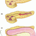
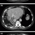
Fig. 9.1
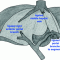
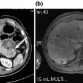
A contrast–enhanced CT showed the metastatic lesions of the right lobe, involving segments 8 and 9, infiltrating the right anterior wall of the adjacent vena cava
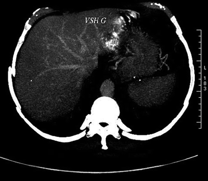
Fig. 9.2
A contrast–enhanced CT showed the second metastatic lesion in the left lobe, in segments 2 and 3 near the root of the left hepatic vein
Because right hepatectomy with en-bloc resection of the involved portion of IVC would have been required for the first lesion, a two-stage procedure was deemed necessary as an initial approach, with subsequent left lateral sectionectomy planned for the second lesion at a second stage.
We will describe here a safe surgical procedure of purely laparoscopic right hepatectomy extended to segment 1 using an anterior approach with partial IVC resection.
Patient Positioning
A low lithotomy, i.e., “French position,” with both the legs abducted at the hip and flexed at the knees and the patient in reverse Trendelenburg position was used (Fig. 9.3). Other routine precautions like adequate padding of the pressure points and thermal covers for the exposed limbs should be followed, and were applied to the case described here.
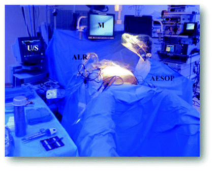

Fig. 9.3
Shows the patient positioning, a low lithotomy, i.e., “French position,” with both the legs abducted at the hip and flexed at the knees and the patient in reverse Trendelenburg position
Trocar Placement
It is difficult to describe a universally acceptable trocar position because of minor variations needed pertaining to each case. Nevertheless, five trocars are usually introduced for standard laparoscopic right hepatectomy. The optical trocar should be located high enough to access the hepatic dome, with two trocars just under the rib, introduced after the establishment of the pneumoperitoneum.
Hand-assist ports can be helpful, especially in case of emergency situations like bleeding, for surgeons with minimal expertise in advanced laparoscopic procedures [13]. In patients with colorectal liver metastases, if there is a previous colostomy then it can be used for gel-ports. In the case described here, the three main working ports were sited subcostally in the right upper quadrant, and a hand-assisted device was inserted at the site of the original colectomy scar, as shown in Fig. 9.4. Apart from use for specimen retrieval, this port proved very useful in the event of major hemorrhage.
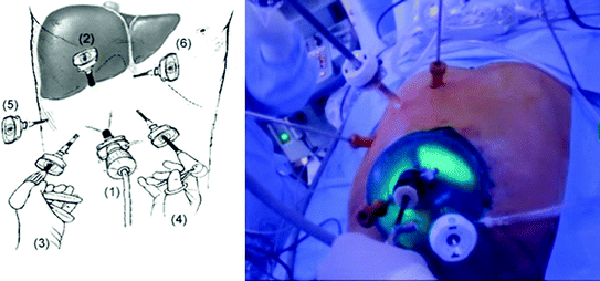

Fig. 9.4
Shows the trocar placement; the three main working ports were sited subcostally in the right upper quadrant and a hand-assisted device was inserted at the site of the original colectomy scar. The illustration shows the trocar placement we use for a right hepatectomy
Surgery
Following insufflation, the abdominal cavity was explored to confirm the absence of disseminated disease. Intra-abdominal pressure was maintained at 12 mmHg. Of note, a higher intra-abdominal pressure was avoided due to a potentially higher risk of gas embolism. A large gauze was placed intra-abdominally, should a compression for haemostasis be necessary in emergency. This can be done through the hand-assist system without losing intra-abdominal pressure. Intraoperative ultrasound was performed to define the extent of the lesion located at the junction of segments 8 and 9 involving IVC. The surgery began with the dissection on the hilum. Cholecystectomy was performed, without removing of the gallbladder, so as to allow a simple way to grasp and retract the liver. Helped by retraction of the cystic duct stump, the hilar region was carefully dissected. The posterior right branches of the bile duct were divided. The hepatic artery and portal vein were isolated and divided between clips, which was usually performed in this order.
Next, the anterior right branches were divided. Hilar dissection was completed to allow access to the IVC. Further dissection was continued in this area by dividing the short hepatic veins and small caudate branches. Here the laparoscopy enabled a very clear view of the anterior surface of IVC from the caudate side. With the line of demarcation established on the liver surface following division of the right hepatic inflow, parenchymal transection via bipolar electrocautery was commenced along Cantlie’s line using an ultrasonic scalpel. The tributaries of the middle vein to segments 5 and 8 were identified, and blood loss was controlled by bipolar electrocautery. Because in this particular case a subsequent left lateral sectionectomy was planned, the middle hepatic vein was not exposed during the right hepatectomy in order to prevent potential injury. The location of the second lesion necessitated the sacrifice of the left hepatic vein, thus leaving the middle hepatic vein as the sole venous outflow of the remnant liver. The parenchymal transection was continued to the hepatic vein/IVC junction. This junction was completely cleared with scissor before the vein was cut using the Endo GIA™ Universal Stapling System.
After the right hepatic vein was divided, the space above the retrohepatic IVC was opened to allow close inspection, in order to delineate whethere there was macroscopic involvement of tumor growth or fibrosis. Partial resection of the IVC was performed by serial applications of the endovascular stapler (Fig. 9.5). Caval resection can be alternatively accomplished with a non-absorbable monofilament whipstitch and vascular clamp. All remaining bands tethering the resected specimen to the diaphragm and retroperitoneum were divided. Haemostasis was verified and the resected margin was carefully inspected for bile leakage, which was controlled with monofilament sutures. The specimen was extracted through the hand-assist system placed on the previous incision.
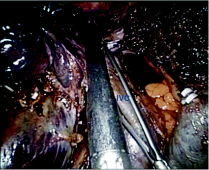

Fig. 9.5
Shows an intraoperative picture of the partial resection of IVC performed by serial applications of the endovascular stapler. A vascular clamp is always ready and under vision intracorporeally when using the stapler devices for hepatic veins or IVC
No drainage was used. The surgical duration was 270 min and the blood loss was 50 ml.
Histological Analysis and Postoperative Course
Histological analysis revealed moderately differentiated adenocarcinoma with a 2 mm surgical margin. The surrounding liver parenchyma showed steatosis. The postoperative period was uneventful and the patient was discharged after 9 days. After a selective portal embolization of segment 2 and 3, the segment 4 showed a volume 515 cc and a full laparoscopic left lateral sectionectomy was performed 7 weeks after the first liver resection. The resection was complete and no adjuvant chemotherapy was used.
Tips and Tricks of the Experienced Surgeon
Specific instrumentation for laparoscopic hepatectomy
The laparoscopic liver surgery requires the following specific hardware (besides the standard equipment of laparoscopic surgery, and open surgery equipment available if conversion is required):
Optical at 0° or 30°, or, better, one flexible 3D optical [14]
Laparoscopic intraoperative ultrasound
Liver retractor
Adjustable cutting linear stapler with vascular several refills
Clips (metal clips, locked clips)
Plastic bag for extraction
To ensure the dissection of the vessels and bile ducts and parenchymal transection, various specific methods can be used (new instruments using different types of energy to dissect the parenchyma, or coagulate vessels in the closing time of hepatic transection) [15].
Coagulator argon should not be recommended in laparoscopy, although some new-generation devices allow a stream of argon at a pressure 10 times lower than the first devices, because of the risk of air embolism [16]. The mechanical fastening clips can be used for elements of the section of the pedicle and the hepatic veins, but also parenchymal transection.
Ultrasound
An important instrument in liver resection is an intraoperative ultrasound [17]. Both in open and laparoscopic liver surgery, ultrasound of the liver is performed for identification of other lesions, to confirm the relationship of the lesion with the portal and hepatic veins [18] and to analyze the distance between the segment 8 vein and IVC.
In laparoscopic approach, until the 3D camera becomes easily available, one of the main risks during the hepatic transection phase is struggling to maintain the correct plane, making intraoperative ultrasonography (IOUS) absolutely mandatory. Although the use of a 3D camera may make this transection phase easier, it will not replace IOUS. Performing a laparoscopic ultrasound of the liver can be a challenging procedure if one has only a rigid device. Thus, a modern ultrasound device should be flexible and should have a guidance system for an RFA-needle.
For right hemi-hepatectomy, IOUS is useful in confirming the position of segment VIII draining vein into either MHV or directly into IVC (Fig. 9.6), which might avoid unnecessary bleeding [17].
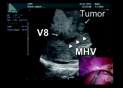

Fig. 9.6
Shows a picture of IOUS showing the position of segment VIII draining vein into either MHV or directly into IVC; in laparoscopic right hepatectomy, the position of this vein is systematically checked before the hepatic transection
Technical Alternatives
If the surgeon is not experienced, we recommend a first manually assisted laparoscopic right hepatectomy. The arguments in favor of hand-assisted technique are the possibility of liver palpation, easier exposure, especially in the first and lower posterior segments and speed and efficiency in control of bleeding [13].
Control of Hemorrhage
In open liver surgery, inflow control (Pringle maneuver) is a tool to decrease blood loss. It is mainly used for major resections. In laparoscopic liver resection, it is feasible to obtain inflow control by performing a similar Pringle maneuver [19]. Although it can be useful, it is not a standard procedure. However, we advise becoming experienced performing a Pringle maneuver, at least in the beginning of laparoscopic liver surgery [13]. After opening the pars flaccida of the lesser omentum, a traumatic forceps or a dissector passed through the hiatus of Winslow and tape is placed around the hepatic pedicle (inserted into a rubber tube for pedicle clamping lakes).
If used, the clamping is preferably intermittent (15 min clamping and 5 min of unclamping). In cases of cirrhotic, steatotic, or blue liver (post chemotherapy), which cannot tolerate prolonged ischaemia, isolated arterial Pringle could be also a useful maneuver [20].
Pneumoperitoneum with an intra-abdominal pressure of 12 mmHg itself will decrease hemorrhage during parenchymal transection, which can also be increased up to 15 mm of Hg if needed [21].
Bipolar devices can be used to stop minor bleeding from the hepatic veins [19]. Vascular and bile structures over 3 mm are controlled by clips, then sectioned.
Stay updated, free articles. Join our Telegram channel

Full access? Get Clinical Tree


