Fig. 1.1
Thyroid development—anterior view. The thyroid originates at the foramen cecum in the midline embryonic pharynx and descends as the thyroid diverticulum to the lower neck to its final position anterior to the trachea. Parafollicular C cells originate from the ultimobranchial bodies adjacent to the 4th pharyngeal pouches and join the developing median thyroid during its migration
Follicles appear within the thyroid gland at the beginning of the second month of gestation and most follicles are formed by the end of the fourth month of gestation. Even though it is the first endocrine gland to form, the foetal thyroid does not produce thyroid hormones until 18–20 weeks of gestation. Therefore, for the first half of gestation the foetus is dependent on maternal thyroid hormones that cross the placenta. This supply of maternal thyroid hormone is especially critical for early neurological development. Maternal hypothyroidism can lead to marked neurodevelopmental compromise of the child [1].
In addition to thyroid hormone-producing follicular cells another group of hormonally active cells in the thyroid are the calcitonin-producing parafollicular C cells. Parafollicular C cells originate from the neuroectoderm of the fifth pharyngeal pouch, the ultimobranchial bodies. They migrate from their lateral origins to merge with the midline developing thyroid. C cells are scattered throughout the mature thyroid gland but are concentrated near the embryologic merger point at the central posterior portion of the upper third of each thyroid lobe.
The molecular genetics of thyroid embryology has been investigated and many transcription factors such as FGF10, FOXE1, HHEX, HOXA3, NKX2-1 AND PAX8 have been identified that are essential for normal thyroid development [2]. For example, heterozygous mutations in PAX8 are associated with thyroid hemiagenesis. Furthermore, gene mutations and copy number gains in key pathways occur in thyroid tumours. Examples of such abnormalities include the RET (rearranged during transfection)-Ras-BRAF-MEK and RET-beta-catenin pathways in medullary thyroid carcinoma in Multiple Endocrine Neoplasia 2A and 2B, FAP (familial adenomatous polyposis) and TRK-PI3K-Akt in papillary thyroid carcinoma, and MDM-p53-PTEN (phosphatase and tensin homolog) pathway in follicular thyroid carcinoma in Cowden syndrome [3]. Another mechanism of papillary thyroid tumourigenesis results from rearrangement of genes encoding receptors with tyrosine kinase activity such as RET rearrangements with H4, PRKAR1A, ELE1, PCM1, RFG5, TIF1A, and TIF1G, known as RET/PTC1-7 and NTRK1 rearrangements with TPM3, TPR and TFG. Autonomous activation of these tyrosine kinase receptors stimulates growth of follicular thyroid and anaplastic thyroid carcinomas via the PI3 K/Akt and MAPK pathways [4]. As a consequence, tyrosine kinase inhibitors are being investigated as a potential treatment for advanced thyroid cancer.
Embryological Anomalies of the Thyroid with Clinical Significance
Thyroid dysgenesis is the term used to describe abnormalities of thyroid gland development and includes a spectrum of conditions including thyroid agenesis (complete absence of thyroid tissue), thyroid hypoplasia (less than the normal amount of thyroid tissue), and thyroid ectopia (thyroid tissue in abnormal locations). Agenesis of the thyroid gland is a very rare anomaly and like thyroid hypoplasia is a cause of congenital hypothyroidism that must be recognized and treated with thyroid hormone replacement in early infancy to prevent neurodevelopmental compromise. One form of hypoplasia is hemiagenesis when one lobe does not develop. In this rare condition the left lobe is more commonly absent.
A thyroglossal duct cyst is a remnant of the diverticulum that forms along the migration path of the thyroid primordium from the foramen caecum at the base of the tongue to the thyroid gland’s final position in front of the trachea. This diverticulum usually begins to involute during the 5th week of development but incomplete involution may result in ectopic thyroid tissue that is typically located in the midline or just off the midline between the hyoid bone and the thyroid isthmus (Fig. 1.2). Thyroglossal duct cyst is the most common congenital neck mass found in children and it can also present later in life in the same location. Thyroglossal duct cysts may enlarge, become infected or rarely undergo malignant transformation. The treatment of a thyroglossal duct cyst is surgical excision of the cyst and (to reduce the chance of recurrence) the entire course of the thyroglossal duct including the middle third of the hyoid bone. This is known as the Sistrunk procedure [5].
Failure of the thyroid migration results in a lingual thyroid which is the most common form of complete thyroid ectopia. Lingual thyroid is a relatively rare condition that is more prevalent in women and in the majority of cases it is associated with an absence of a normal cervical thyroid. It is generally presents incidentally as an asymptomatic mass at the back of the tongue (Fig. 1.2). If large, a lingual thyroid can produce obstructive symptoms such as dysphagia, dysphonia, or dyspnoea. When a lingual thyroid is suspected based by clinical findings the diagnosis can be confirmed by radioisotope scanning which also confirms the absence of other thyroid tissue in the neck.
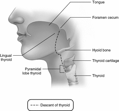

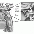
Fig. 1.2
Thyroid development—midline sagittal view of the path of thyroid descent in mature human. Ectopic thyroid can be present anywhere along the path such as in the midline posterior tongue where it is known as a lingual thyroid or attached to the thyroid isthmus as the pyramidal lobe. Remnants of the thyroid diverticulum known as thyroglossal duct cysts or sinuses can occur anywhere along the path, most commonly in the midline neck between the hyoid bone and thyroid
Thyrothymic thyroid rests are ectopic collections of thyroid follicular cells often connected to the lower lobes of the thyroid but sometimes completely separate. In either case, they descend in the anterior or posterior mediastinum and may cause symptoms if they enlarge. This abnormally located thyroid tissue can present similarly to the more common retrosternal goitre which is an exuberant growth of normally located thyroid lobes down into the mediastinum.
Struma ovarii, also known as thyroid goitre of the ovary, is an ovarian teratoma that contains mostly thyroid tissue. The teratoma is a developmental anomaly of foetal gonadal tissue. This exceedingly rare condition is usually an unexpected histological finding in patients who have excision of an ovarian mass.
Embryology of the Recurrent Laryngeal Nerve
The recurrent laryngeal nerve (RLN) is a branch of the vagus nerve going to the larynx and its final path is linked to developmental changes in the aortic arches. By the 6th–7th week, the lowest persisting aortic arch on each side pulls the respective RLN downwards. On the left, the RLN passes around the 6th aortic arch which in the foetus becomes the ductus arteriosus that normally closes postnatally and becomes the ligamentum arteriosum. It then travels upwards in the tracheo-oesophageal groove. On the right, the 5th and 6th aortic arches disappear and the RLN passes around the 4th aortic arch which becomes the right subclavian artery. From this position the right RLN then ascends more obliquely than the left RLN (Fig. 1.3). As a consequence, the left RLN is longer than the right RLN and, during intraoperative nerve monitoring there is a slightly longer latency of the signal obtained during stimulation of the vagus on the left side compared with the right side.
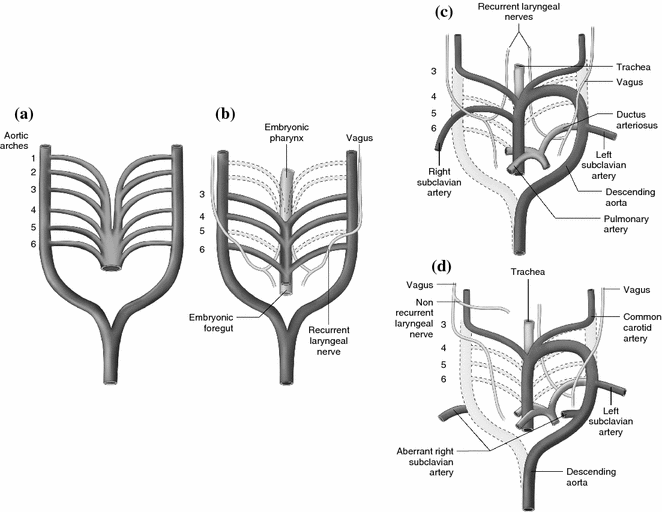

Fig. 1.3
Aortic arch development and recurrent laryngeal nerve. a Embryonic paired aortic arch vessels, also known as pharyngeal arch arteries correlating to developing pharyngeal arches. b Embryonic paired aortic arches with recurrent laryngeal nerve (RLN). The RLN loops around the lowest (6th) aortic arch on each and then goes to the larynx. In this depiction the 1st, 2nd, and 5th aortic arches have already regressed. c As normal development proceeds the right 6th aortic arch regresses so that the right RLN loops around the lowest remaining arch, the 4th aortic arch which becomes the right subclavian. The left RLN passes around the persisting 6th aortic arch which in the foetus becomes the ductus arteriosus and postnatally normally closes to becomes the ligamentum arteriosum. d When the right 4th aortic arch does not develop then an aberrant right subclavian artery originates from the aortic arch distal to the right carotid, left carotid and left subclavian artery and passes to the right upwards and behind the oesophagus. With no embryological right aortic arch to pull it downward, the right RLN nerve goes straight from the vagus into the larynx and is therefore “non-recurrent”
An embryological vascular anomaly (present in 0.5–1% of people) leads to a non-recurrent laryngeal nerve and illustrates the close connection between the developing RLN and aortic arches. When the 4th aortic arch on the right does not develop an aberrant right subclavian artery originates from the left side of the aortic arch, distal to the origins of the right carotid, left carotid and left subclavian artery. From this origin it passes upwards and to the right behind the oesophagus. Occasionally this retro-oesophageal location is associated with esophageal compression and dysphagia explaining its designation as arteria lusoria. Since there is no embryological right aortic arch to pull the right RLN downwards the nerve goes straight from the vagus into the larynx and is therefore “non-recurrent” (Fig. 1.3). This unusual vascular anomaly can be demonstrated on Doppler ultrasound, which in some centres is part of routine preoperative investigation before thyroid surgery. The use of intraoperative neuromonitoring may also identify patients with this anatomical anomaly [6] when stimulation of the distal vagus nerve does not produce a RLN signal while stimulation proximal to the origin of the non-recurrent RLN generates the correct signal [7]. The diagnosis can be confirmed postoperatively by ultrasound identification of the vascular anomaly or by a barium swallow showing the retroesophageal artery as an extrinsic compression of the posterior oesophagus. On the left side the non-recurrent RLN is exceptionally rare with only a few cases reported. A more common finding is a false non-recurrent RLN which is seen when a large anastomotic branch of the cervical sympathetic chain joins a normal RLN.
Anatomy of the Thyroid Gland
Gross Anatomy
The normal adult thyroid weights 20–30 g and has two lobes measuring approximately 5 cm in length and 3 cm in width. The thyroid gland lies between the anterior borders of the sternocleidomastoid (SCM) muscles in the anterior triangle of the neck in front of the trachea caudal to the cricothyroid membrane and the thyroid cartilage (“Adam’s apple”), with the isthmus overlying the second to fourth tracheal rings. A pyramidal lobe can be observed in some 50% of patients as an extension of thyroid tissue from the isthmus towards the hyoid bone (Fig. 1.4).
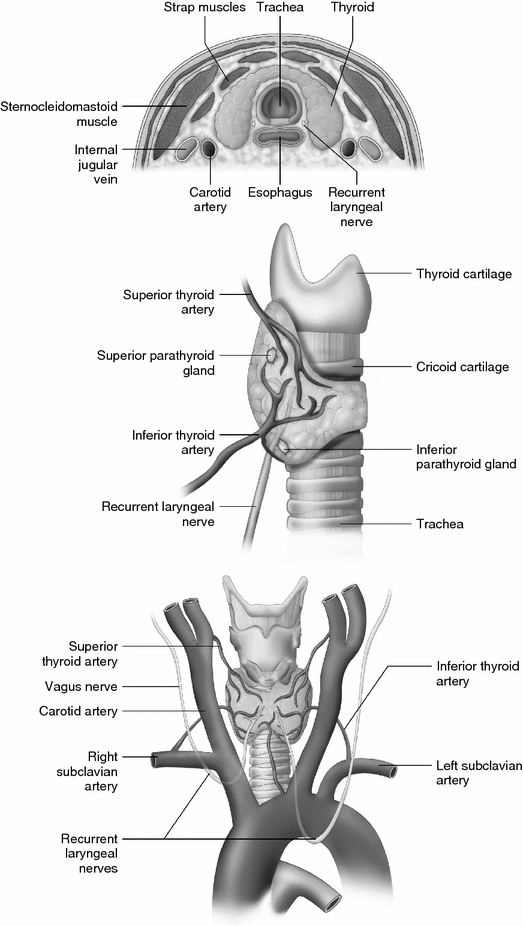

Fig. 1.4
Thyroid anatomy. Anterior, lateral, and cross-sectional
A midline mass situated at or above the thyroid cartilage is usually not the thyroid gland but more likely a thyroglossal cyst or an enlarged lymph node. A mass visible along the edge or just under the SCM is more likely to be a branchial cyst. A mass visible lateral to SCM (i.e. in the posterior triangle) is more likely to be an enlarged lymph node.
Surgical Anatomy of Classical Thyroidectomy (in the Order Encountered During the Operation)
- 1.
The skin incision follows Langer’s lines or the wrinkle lines in the neck overlying the thyroid isthmus. Kocher was the first to describe the transverse collar incision [8].
- 2.
The subcutaneous fat contains numerous small blood vessels.
- 3.
The platysma muscle is a broad and very thin muscle layer extending from the deep fascia of the upper pectoralis muscle to the mandible and lower face. The platysma muscle may be incomplete with bands of muscle separated by subcutaneous fat in the lower part of the neck and therefore might not be seen with short incisions. Immediately beneath the platysma are the anterior jugular veins.
Stay updated, free articles. Join our Telegram channel

Full access? Get Clinical Tree


