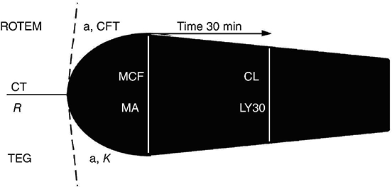
Chapter 27 Jon Bailey1 and Nicola S. Curry2,3 1 Oxford University Hospitals, John Radcliffe Hospital, Oxford, UK 2 Department of Haematology, Oxford University Hospitals, Oxford University, Oxford, UK 3 Oxford Haemophilia and Thrombosis Centre, Oxford University Hospitals, Oxford, UK Coagulopathy (hypo- and hypercoagulability) is common in the intensive care setting. Traditionally, coagulation potential has been assessed using plasma-based tests, such as the prothrombin time (PT) or the activated partial thromboplastin time (APTT); but these tests have repeatedly been shown to be poor predictors of bleeding and thrombosis [1]. Reliable prediction and/or diagnosis of clinically relevant clotting dysfunction and subsequent monitoring of effective treatment are important management issues in critically ill patients. Viscoelastic tests, such as TEG® or ROTEM®, are increasingly being used to predict coagulopathy and guide transfusion therapy. This chapter will briefly describe the mechanics of these tests, discuss their potential for use in the critical care setting and touch on the evidence that supports their use. Both TEG® (Haemonetics Corp., Braintree, MA, USA) and ROTEM® (Tem International GmbH, Munich, Germany) are viscoelastic haemostatic assay (VHA) devices which assess the global elastic properties of clot formation under low shear stress. Whole blood (native or citrated) is used, and the interaction of coagulation factors, and their inhibitors, with red blood cells and platelets during clotting and fibrinolysis is evaluated, usually over 60 min. Both devices measure the speed of clot formation, the strength and stability of the clot when formed and the kinetics of clot breakdown (fibrinolysis) (see Figure 27.1 and Table 27.1). VHAs are argued to be better measures of overall haemostatic potential than plasma-based tests, providing a more physiologically relevant interpretation of coagulation. However, the results from the two devices, although similar, should not be regarded as directly comparable since different coagulation activators are used (see Table 27.2). Figure 27.1 Diagram of a typical TEG®/ROTEM® trace. Table 27.1 TEG and ROTEM variables. A mathematical formula determined by the manufacturer can be used to determine a coagulation index (CI) that takes into account the relative contribution of each of r, K, α and MA into a single complex equation. Table 27.2 Viscoelastic Haemostatic Assays.
The Relevance of Thromboelastography in Intensive Care Patients
Viscoelastic tests
Process studied
Causes of variation
TEG value
ROTEM value
Time until first evidence of clot formation (amplitude of 2 mm reached)
Prolonged by clotting factor deficiencies and heparin
R value (reaction time)
Clotting time (CT)
Rate of clot formation (time for amplitude to increase from 2 to 20 mm)
Decreased by clotting factor deficiencies such as hypofibrinogenaemia and platelet dysfunction or insufficiency
K value and α angle
α angle and clot formation time (CFT
Maximum clot strength
Reduced by platelet dysfunction and hypofibrinogenaemia
Maximum amplitude (MA)
Maximum clot firmness (MCF)
Clot lysis (CL)
Measures degree of fibrinolysis
LY30 – CL at 30 min
CL
TEG
ROTEM
Test
Activator
Test
Activator
Diagnostic use
NATEM (native TEG)
Coagulation without added activator
Kaolin-activated TEG
Kaolin
INTEM
Ellagic acid
Defects in the intrinsic pathway of coagulation activation, heparin anticoagulation
EXTEM
Recombinant TF
Defects in the extrinsic pathway, prothrombin complex deficiency, platelet deficiency
RapidTEG
Kaolin and TF
Defects in the intrinsic and extrinsic pathways
FF reagent
TF and abciximab
FIBTEM
Cytochalasin D
Fibrin-based clot defects, fibrin/fibrinogen deficiency
Can help differentiate low fibrinogen values and platelet dysfunction which both cause low EXTEM MCF or low MA
APTEM
Aprotinin
Evaluates fibrinolysis, when compared to EXTEM values
Kaolin-activated TEG + heparinase
Kaolin and heparinase
HEPTEM
Heparinase
Heparin/protamine imbalance
Related posts:


Stay updated, free articles. Join our Telegram channel

Full access? Get Clinical Tree


