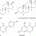
Fig. 13.1
Plasma ratio (40:1) and pharmacokinetics of MI and DCI. Data of two different studies in human volunteers
Recently, three different studies evaluated the efficacy of a treatment based on the physiological plasma ratio between MI and DCI of 40:1 in PCOS women. The idea behind this therapy is that an INS dysregulation plays a central role in nurturing PCOS. Indeed, epimerase dysregulation changes the MI:DCI ratio, which in turn could impair hormone signaling, namely, of both insulin and FSH.
Various evidences supported a deficiency concerning the availability and/or utilization of MI and/or DCI in tissues of PCOS women, and this impairment likely contributes to the insulin resistance typical of that syndrome [6, 10]. Unlike other tissues, such as muscles and liver, the ovaries are not insulin resistant. Because the epimerase activity, regulating the MI:DCI ratio, is insulin dependent, PCOS patients are affected by a boosted MI to DCI epimerization into the ovary, leading to overproduction of DCI and MI deficiency [11], as shown by two independent laboratories [6, 10]. Thus, a specific MI depletion and a DCI overload characterize the ovary of PCOS women. The poor oocyte quality observed in PCOS patients can be explained by this imbalance, responsible also for the impaired FSH signaling [12, 13].
Literature evidences have already shown that MI supplementation is able to correct PCOS metabolic aspects. Two trials demonstrated that the same effect was obtained even in a more effective way by administering MI and DCI in a physiological ratio (40:1). Indeed, the improved parameters were diastolic blood pressure, fasting glucose, fasting insulin, and both insulin and glucose AUCs [14, 15]. Additional improved parameters were those linked to the CVD, namely, HOMA index, triglycerides, and both HDL and LDL cholesterol. Noteworthy, ovulation was restored in the majority of the women.
Furthermore, by moving from the metabolic aspects of the syndrome to the reproductive ones, a trial has shown that the treatment of PCOS women undergoing ICSI, with a MI:DCI 40:1 based therapy, retains the beneficial effects of MI treatment alone, outperforming the DCI treatment [12].
In particular, the treatment is able to improve ovarian response and oocyte and embryo quality. Recently, the interest of the scientific world on MI and DCI has pushed the PRESIS to organize an international consensus conference in order to clarify this issue and lay the foundations of future researches.
13.4 Conference Aim and Methods
Since the knowledge of the differences between MI and DCI is not well established among researchers, as it is proven by a systematic and a Cochrane review mixing trials performed using MI or DCI, the PREIS School (Permanent International and European School in Perinatal Neonatal and Reproductive Medicine) has organized the “2013 Florence International Consensus Conference on Myo and d-chiro-inositol in Obstetrics and Gynecology and Assisted Reproduction Technology (ART)” aimed at elucidating some controversial points with the contribution of opinion leaders in the fields of cell biology, mammalian embryology, human endocrinology, metabolism, obstetrics, and gynecology. Two separate panels of this Committee worked on the roles of MI:DCI in metabolic syndrome (mainly PCOS) therapy and of MI in ART and drew up two lists of hot topics. Our review reports only the published results in the paper on myo-inositol and ART [16].
The following is a set of research questions concerning ART:
1.
Physiological involvement of INS in oocyte maturation
2.
INS involvement in the physiology of spermatozoa function
3.
Usefulness of the treatment with INS during ART cycles
4.
Comparison of the clinical efficacy between supplementation with MI and/or DCI
13.5 MI and ART
13.5.1 Physiological Involvement of INS in Oocyte Maturation
13.5.1.1 Role of MI in Oogenesis and Early Embryogenesis
In mammalian females including humans, an elevated MI content in the follicular fluid fosters oocyte quality and pregnancy outcome [17, 18]. MI activity is in connection with the InsP3 function on the modulation of intracellular calcium ion concentration, influenced by LH and FSH hormones [16]. In oocytes, MI, among different functions at the ovarian level, positively affects the maturation process [16]. The decrease of intracellular MI stores impairs oocyte maturation, and MI supplementation in culture medium has been shown to increase the development of fertile eggs [16]. The implantation rate and post-implantation viability of embryos rise if the oocytes are cultured in a medium containing MI and then fertilized in vitro and transferred for promoting pregnancy [16–19]. During in vitro fertilization (IVF) cycles, the treatment of women with MI before the hormonal stimulation has reduced the FSH quantity to be administered and the number of days required for the appropriate stimulation. All these parameters are positively related to the possibility of pregnancy and improved quality of oocytes and embryos and, probably, the implantation rate [12, 16]. Thus, MI administered 3 months before ovulation induction can produce a rise in the number of high-quality embryos obtained in IVF cycles.
13.5.1.2 MI and Oogenesis: A Lesson from Polycystic Ovary Syndrome
Further proofs confirming the essential role of MI in follicular fluid for safeguarding egg quality derived from the PCOS studies were previously examined. It is therefore clear that MI depletion in a PCOS ovary impairs dominant follicle recruitment and appropriate oocyte growth/maturation. These data support the fundamental observations by Chiu et al. [18] showing that proper content of MI in follicular fluid indicates a required condition to ensure egg quality.
13.5.2 INS Involvement in the Physiology of Spermatozoa Function
In agreement with the MI high levels in female generative system, the same condition can be found in mammalian male, where MI content is more elevated in reproductive organs than in blood serum and increases from the caput to the cauda epididymis [16]. In males, FSH-responsive Sertoli cells are the main producers of MI, which is implicated in processes such as the regulation of spermatozoa motility, capacitation, and acrosome reaction. MI increases sperm cell parameters in male patients suffering from oligoasthenoteratozoospermia (OAT), a severe pathology impairing sperm cell number, morphology, and function [16]. This evidence suggests that MI use in the treatment of semen samples during IVF cycles can raise fertilization rate and embryo quality, in this way, giving higher chances of pregnancy. Treating OAT patients’ sperm cells with MI gives the following changes: the presence of amorphous material and semen viscosity decreases, midpiece volume improves, and mitochondrial cristae morphology is restored, regularizing the mitochondria structures [16]. At the functional level, a key step is the direct MI action on mitochondria, raising the membrane potential [16]. High values of mitochondrial membrane potential attest to the integrity of this structure, meaning, optimal levels of activity and proper cell viability. Therefore, MI treatment of sperm cells from both OAT patients and normal subjects enhances the recovery of cells usable in IVF cycles after swim-up [16], supporting its use as supplement in sperm cells manipulation in the procedures of medical-assisted reproduction.
Stay updated, free articles. Join our Telegram channel

Full access? Get Clinical Tree


