The Process of Pain in the Cancer Patient
“Pain is an unpleasant sensory and emotional experience associated with actual or potential tissue damage, or described in terms of such damage.”1 The sensory features and affective qualities of pain vary, depending on its origin, the previous experiences of the patient, and the environment that surrounds the patient. Its negative affective characteristics are related to the social and physical context in which pain occurs, as well as to cognitive processes such as the meaning of tissue trauma for the individual.
SENSORY COMPONENT OF CANCER PAIN
Tumor-associated pain is either nociceptive or neuropathic, or a combination of the two. Nociceptive pain is thought to be related to unhealed tissue injury of either somatic or visceral structures. Pain associated with injury to neural tissues is sustained by aberrant somatosensory processing in the peripheral or the central nervous system; it is labeled as neuropathic. An international survey of cancer pain characteristics and syndromes reported that pains thought to be nociceptive and caused by somatic injury occurred in 71.6% of patients, nociceptive visceral pain in 34.7%, and neuropathic pain in 39.7%.2 Somatic nociceptive pain may be divided into superficial (cutaneous) and deep. Cutaneous pain usually is well localized, sharp, pricking, or burning. Deep tissue pain is usually reported as diffuse and dull or aching in quality. Visceral pain is usually poorly localized, often referred to the body surface, of long duration, and commonly described as “sickening.”
Tumor involvement of the peripheral nervous system has many manifestations and can include lesions within the cerebrospinal fluid space, local invasion, compression of nerves, direct infiltration, perineural spread, and intraneural metastasis, although the last is quite rare.3 Both myelinated and unmyelinated fibers and supporting tissues are destroyed by compression or invasion of nerves by tumor. Infiltration by a neoplasm is the prototypic tissue trauma stimulus. A remote neoplasm may produce indirect damage of unknown pathogenesis in peripheral nerves (eg, paraneoplastic syndrome). Tumor may also directly invade nerve.4 Primary neural tumors also can lead to painful destruction of the parent nerve. Neural compression and the associated inflammatory changes can also lead to pain, especially when the nerve is confined by osseous structures, as, for example, where the dorsal root goes through the neural foramen.
Degenerative, regenerative, and other pathophysiologic processes are involved when a nerve is compressed or invaded. 5 The entire afferent neuron, dendrites, soma, and axon manifest biochemical changes that are both reactive and reparative. Eventually, the neuron loses its neuropeptides,6 atrophies, and degenerates. Unmyelinated afferent neurons are particularly susceptible to this process. Neural degeneration after invasion or compression by cancerous tissue is probably similar to that seen after lesions of nerves induced by mechanical or other events. Changes occur not only in impulse transmission but also in the resting activity, as well as in responses to mechanical, thermal, and chemical stimulation.
SENSITIZATION OF NOCICEPTORS
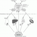
Neoplasms contain, in addition to cancer cells, different cell types, such as inflammatory and neo-vascular cells, that are in proximity to primary afferent nociceptors (Fig. 4.1). Most of these cells belong to the immune system, including macrophages, neutrophils, and T-lymphocytes known to secrete various factors that sensitize or directly excite primary afferent neurons. These factors include prostaglandins, tumor necrosis factor (TNF), endothelins, interleukin-1 and -6, epidermal growth factor, transforming growth factor, and platelet-derived growth factor. Primary afferent neurons express receptors for many of these factors. Endothelins are a family of vasoactive peptides that are expressed at high levels by several types of tumor, including prostate cancer. Endothelins contribute directly to cancer pain by sensitizing or exciting nociceptors, because a subset of small, unmyelinated primary afferent neurons expresses endothelin-A receptors.7 Angiogenesis and tumor growth are thought to be regulated by endothelins and prostaglandins that are produced by cancer cells.8 These and many other factors, as well as others produced by cancer and inflammatory cells such as adenosine triphosphate (ATP), bradykinin, H+, nerve growth factor (NGF), prostaglandins, and vascular endothelial growth factor (VEGF) sensitize or even excite adjacent nociceptors.9 Endothelins released by cancer cells are detected by the nociceptor vanilloid receptor-1 (VR1), which is also sensitive to extracellular H+. The tyrosine kinase receptor TrkA binds to NGF that has been released by macrophages.10 Neurotransmitters, such as calcitonin gene-related peptide (CGRP), endothelin, histamine, glutamate, and substance P are liberated during nociceptor activation. Prostaglandins released from the peripheral terminals of sensory fibers during their activation can lead to vasodilatation, plasma extravasation, and the recruitment and activation of immune cells.
Tumor-related pain can be induced by tissue acidosis and other mechanisms. Decreases in tissue pH can be explained via several mechanisms. When inflammatory cells invade neoplastic tissues, they release protons that
generate local acidosis. Apoptosis commonly occurs in the tumor environment and contributes to acidosis, as apoptotic cells release intracellular ions, creating an acidic environment. The fall in pH can activate signaling by acid-sensing channels (including VR1) expressed on nociceptors. The generation of bone cancer pain may be dependent upon the tumor-induced release of protons and acidosis.11 Tumor invasion of sensory fibers may also lead to pain, although tumors are not significantly innervated by sensory neurons.12 However, rapid tumor growth may entrap and injure nerves, producing mechanical injury, compression, ischemia, or direct proteolysis. Tumor cells can also produce proteolytic enzymes that can cause injury to sensory and sympathetic fibers, leading to a neuropathic pain syndrome.
generate local acidosis. Apoptosis commonly occurs in the tumor environment and contributes to acidosis, as apoptotic cells release intracellular ions, creating an acidic environment. The fall in pH can activate signaling by acid-sensing channels (including VR1) expressed on nociceptors. The generation of bone cancer pain may be dependent upon the tumor-induced release of protons and acidosis.11 Tumor invasion of sensory fibers may also lead to pain, although tumors are not significantly innervated by sensory neurons.12 However, rapid tumor growth may entrap and injure nerves, producing mechanical injury, compression, ischemia, or direct proteolysis. Tumor cells can also produce proteolytic enzymes that can cause injury to sensory and sympathetic fibers, leading to a neuropathic pain syndrome.
 FIGURE 4.1 Detection by sensory neurons of noxious stimuli produced by tumors (A). Nociceptors (pink) use different receptors to detect and transmit signals about noxious stimuli produced by cancer cells (yellow) or other aspects of the tumor microenvironment. Vanilloid receptor-1 (VR1) detects H+ produced by cancer cells, whereas endothelial-A receptors (ETAR) detect endothelins (ET). Dorsal-root acid-sensing ion channel (DRASIC) detects mechanical stimuli as tumor growth distends sensory fibers. Other receptors expressed by sensory neurons include prostaglandin (PG) receptors, which detect PGE2 (produced by cancer and inflammatory cells). Nerve growth factor (NGF) released by macrophages binds to tyrosine kinase receptor (TrkA). B: Tumor-nociceptor interface. In addition to cancer cells, tumors consist of inflammatory cells and blood vessels and are often adjacent to primary afferent nociceptors. Cancer cells and inflammatory cells release a variety of products, such as adenosine triphosphate (ATP), bradykinin, H+, NGF, PG, and vascular endothelial growth factor (VEGF), that either excite or sensitize the nociceptor. Painful stimuli are detected by the nociceptors, the cell bodies of which lie in the dorsal root ganglion (DRG), and are transmitted to neurons in the spinal cord. The signal is then transmitted to higher centers of the brain. Cancer-associated pain signals seem to ascend to the brain by at least two main spinal-cord pathways, the spinothalamic tract and the dorsal column. Nociceptor activation results in the release of neurotransmitters, such as calcitonin gene-related peptide (CGRP), endothelin, histamine, glutamate, and substance P. Nociceptor activation also causes the release of PGs from the peripheral terminals of sensory fibers, which can induce plasma extravasation, recruitment and activation of immune cells, and vasodilatation.9 VR1, Vanilloid receptor-1; ETAR, endothelial-A receptor; ET, endothelin; DRASIC, dorsal-root acid-sensing ion channel; PG, prostaglandin; NGF, nerve growth factor; TrkA, tyrosine kinase receptor; ATP, adenosine triphosphate; VEGF, vascular endothelial growth factor; DRG, dorsal root ganglion; CGRP, calcitonin gene-related peptide. (Adapted from Mantyh PW et al. Molecular mechanisms of cancer pain. Nat Rev Cancer 2002;2(3):201-209. Copyright 2002, with permission from Macmillan Publishers, Ltd.) |
CAUSES OF PAIN IN THE CANCER PATIENT
Cancer pain is a mixture of acute pain, chronic pain, tumor-specific pain, and treatment-related pain, all compounded by ongoing psychological responses of distress and suffering. Almost all pain in cancer patients may be considered the result of one or more of three fundamental causes (Table 4.1): direct tumor involvement, cancerdirected therapy, and mechanisms unrelated to cancer or its treatment.
Complex patterns of pain that result from combinations of these categories are common in cancer patients; accurate diagnosis can be difficult. In addition, many elements that contribute to the challenge of controlling cancer-related and chronic noncancer pain (central sensitization, hyperalgesia, novel gene expression, synaptic remodeling, and behavioral adjustment) emerge promptly on persistent tissue injury.13, 14 Factors influencing the pain complaint include the primary tumor type, stage of disease, tumor site, and mood factors (anxiety and depression).15, 16, 17 Vainio et al.17 estimated the prevalence of pain in 1,840 patients with advanced cancer from seven hospices in Europe, the United States, and Australia. Twenty-four
percent of the patients surveyed had no pain, 24% had mild pain, 30% had moderate pain, and 21% had severe pain. In addition, the prevalence of moderate or severe pain was highest in gynecological cancer and in head and neck cancer. Severe pain was most common in prostate cancer (Table 4.2). Van den Beuken-van Everdingen et al.,18 in a meta-analysis of 52 studies, calculated the pooled prevalence of pain in oncology patients. The prevalence of pain in patients after curative treatment was 33%; in patients under anticancer treatment, 59%; in patients with advanced/metastatic/terminal disease, 64%; in patients at all disease stages, 53%. Of the patients with pain, more than one third graded their pain as moderate or severe. Pooled prevalence of pain was >50% in all cancer types, with the highest prevalence in head/neck cancer patients (70%).
percent of the patients surveyed had no pain, 24% had mild pain, 30% had moderate pain, and 21% had severe pain. In addition, the prevalence of moderate or severe pain was highest in gynecological cancer and in head and neck cancer. Severe pain was most common in prostate cancer (Table 4.2). Van den Beuken-van Everdingen et al.,18 in a meta-analysis of 52 studies, calculated the pooled prevalence of pain in oncology patients. The prevalence of pain in patients after curative treatment was 33%; in patients under anticancer treatment, 59%; in patients with advanced/metastatic/terminal disease, 64%; in patients at all disease stages, 53%. Of the patients with pain, more than one third graded their pain as moderate or severe. Pooled prevalence of pain was >50% in all cancer types, with the highest prevalence in head/neck cancer patients (70%).
TABLE 4.1 CAUSES OF PAIN IN ONCOLOGY PATIENTS | ||||||||||||||||||||||||
|---|---|---|---|---|---|---|---|---|---|---|---|---|---|---|---|---|---|---|---|---|---|---|---|---|
|
TABLE 4.2 PREVALENCE OF PAIN (PERCENTAGE OF PATIENTS), BY PRIMARY SITE AMONG PALLIATIVE-CARE CANCER PATIENTS | ||||||||||||||||||||||||||||||||||||||||||||||||
|---|---|---|---|---|---|---|---|---|---|---|---|---|---|---|---|---|---|---|---|---|---|---|---|---|---|---|---|---|---|---|---|---|---|---|---|---|---|---|---|---|---|---|---|---|---|---|---|---|
| ||||||||||||||||||||||||||||||||||||||||||||||||
