1 The Normal Breast and Benign Diseases of the Breast
The Normal Breast
The breast is a modified, specialized apocrine gland located in the superficial fascia of the anterior chest wall (Fig. 1-1). The nipple projects from the anterior surface and is hyperpigmented. It is composed of dense fibrous tissue covered by skin and contains bundles of smooth muscle fibers, which assist with milk expression. The skin adjacent to the nipple is also hyperpigmented and is called the areola.
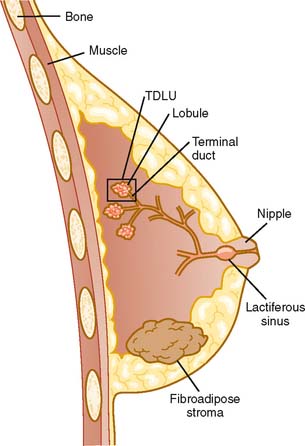
Figure 1-1 Normal breast. Diagram of breast composition and location. TDLU, terminal ductal lobular unit.
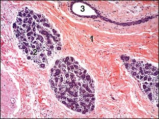
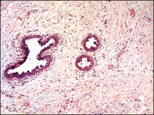
The breast parenchyma consists of 15 to 20 lobules, which drain secretions into a ductal system that converges and opens into the nipple.1 The functional unit of the breast is the terminal duct lobular unit (TDLU) (Fig. 1-2), which is composed of the terminal (intralobular) duct, and its ductules/acini (also referred to as lobules). The terminal ducts join together to form the larger ducts, which have a dilatation (lactiferous sinus) just before they open into the nipple. The TDLUs are embedded in loose specialized, hormonally responsive connective tissue stroma, the intralobular stroma. The dense fibrous tissue between the breast lobules is called interlobular stroma, which is not responsive to hormones (Fig. 1-3).
Lymphatic drainage of the breast is to the axillary, supraclavicular, and mediastinal lymph nodes.
Histology
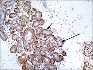
The entire ductal system, extralobular large and intermediate ducts, as well as the intralobular (terminal ducts and ductules/lobules), is lined by two cell layers: inner epithelial cells and an outer interrupted layer of myoepithelial cells.1 The latter cells have contractile properties and assist in expelling milk. Special techniques can be used to highlight the myoepithelial cells. Immunohistochemical stains for muscle-specific actin (MSA), S100, p63, and calponin can be used to detect the myoepithelial cell layer (Fig. 1-4).
Physiologic Changes in Female Breast Histology
Menstrual Cycle
The following are pre- and postmenstrual phases of the menstrual cycle:
Follicular phase: During the follicular phase of the menstrual cycle, the TDLUs are at rest and do not show any growth. The intralobular stroma is dense and indistinct from the dense interlobular stroma.
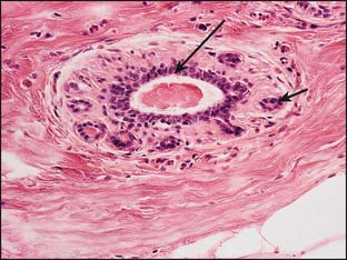
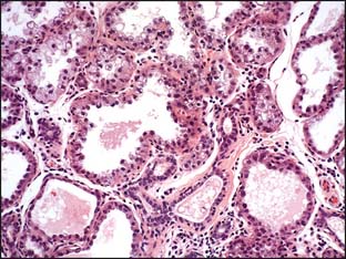
Lactation: In the lactating breast, the individual terminal ducts form acini, which show epithelial vacuolization as a result of the presence of secretions that also fill their lumina (Fig. 1-5). After lactation, the units involute and return to their old structure.
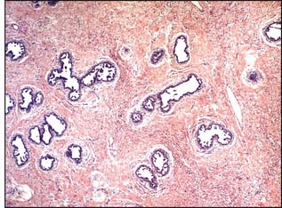
The normal male breast differs in structure from the female breast in that there are no lobules. The male breast consists of ductal structures surrounded by fibroadipose tissue (Fig. 1-6).
Abnormalities of Breast Development
The following are abnormalities that can occur in breast development:
Mammary heterotopia (accessory breasts or nipples) may occur anywhere along embryonic mammary ridges, the most common sites being the chest wall, axilla, and vulva. It may manifest as polythelia (supernumerary nipples) or polymastia (aberrant breast tissue).2 The accessory breast tissue responds to hormonal changes and, if located in the axilla, it may enlarge and raise concern for metastases.
Hamartoma is a well–circumscribed, often encapsulated mass composed of varying combinations of benign epithelial and stromal elements including fat.3 Hamartoma is usually asymptomatic. It may manifest as a palpable mass, or it may be detected by mammography. Hamartoma may cause breast deformity if it is very large.
Gynecomastia
Gynecomastia is defined as enlargement of one or both breasts in a male.4 Many cases are idiopathic. In some cases, gynecomastia may be caused by excessive estrogen stimulation. Predisposing factors include the following:
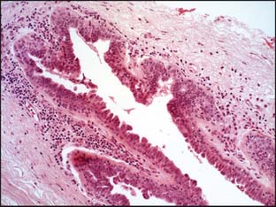
On palpation, a firm disc-shaped subareolar mass is noted. Microscopic features of gynecomastia include ductal epithelial hyperplasia, stromal edema, and fibrosis around ducts (Fig. 1-7).
Inflammatory and Reactive Breast Lesions
The following are inflammatory and reactive breast conditions of various causes:
Acute inflammation of the breast (acute mastitis) is associated with redness, swelling, pain, and tenderness and may occur during the early postpartum months as a result of lactation (puerperal mastitis).5 Staphylococcus aureus is the most common infecting agent. There are two general categories of predisposing factors:
 Stress and sleep deprivation, which may lower the immune status and cause engorgement by inhibiting milk flow
Inflammatory breast carcinoma should be ruled out when there is no response to antibiotic therapy.
Chronic mastitis may be idiopathic6–8 or in response to infection (tuberculosis), foreign material (silicone), or systemic disease (sarcoidosis). Diagnosis requires microbiologic, immunologic, and histologic evaluation. Idiopathic granulomatous mastitis7 is diagnosed after exclusion of specific etiologic agents. Microscopically, chronic mastitis shows granulomas with or without caseation. Surgical excision may be followed by recurrence, abscess formation, or fistula formation.
Mammary duct ectasia is a distinct entity that usually occurs in perimenopausal women as a result of obstruction of the lactiferous ducts by inspissated luminal secretions. Obstruction leads to dilatation of the ducts and periductal chronic inflammation (Fig. 1-8). Grossly, chronic mastitis may produce irregular masses with induration that closely mimic breast carcinoma, and biopsy may be required to exclude carcinoma.
Stay updated, free articles. Join our Telegram channel

Full access? Get Clinical Tree


