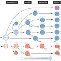
This large and important set of genes owes its rather clumsy-sounding name to the fact that the proteins it codes for were first detected by their effect on transplant rejection, i.e. tissue incompatibility. However, it is now clear that their real purpose is to act as receptors binding and stabilizing fragments of antigen and displaying them at the cell surface for T lymphocytes to recognize, via their own receptors, and activate their adaptive immunological functions.
Again, for historical reasons, the MHC in the mouse (extreme bottom line in the figure) is known as H2, while in humans it is called HLA (human leucocyte antigen). In fact the basic layout of the MHC genes is remarkably similar in all animals so far studied, consisting of a set of class I (shaded in the figure) and a set of class II genes, differing slightly in structure and in the way they interact with T cells (see Fig. 12). In the figure the names of genes are shown boxed, while the numbers below indicate the approximate number of alternative versions or alleles that can occur at each locus. Perhaps the most striking feature of the MHC is the number of different variants that exist in the human population. The number of possible combinations on a single chromosome probably exceeds 3 × 10⁶, so that an individual, with a set of MHC molecules coded for by both chromosomes, can have any one of about 1013 combinations, which is part of the problem in transplanting kidneys, etc. (see Fig. 39).
Since HLA typing became a routine procedure, it has emerged that many diseases are significantly more common, or sometimes rarer, in people of a particular HLA type. There are several mechanisms that might account for this but none of them has yet been established to everybody’s satisfaction.
Peptide-Binding Cleft
The classic MHC I and II molecules contain a peptide-binding site at the distal end of the molecule from the membrane, formed by two protein α-helices, lying on top of a β-pleated sheet. The binding site, or groove as it is often known, can accommodate a peptide of about 9–10 amino acids in length, although for class II MHC molecules, the ends of the groove are open allowing longer peptides to extend out of either end. A wide variety of different peptides can be bound tightly, by interaction between conserved residues in the MHC molecules and the amino acid backbone of the antigen peptide. In order to accommodate the side-chains of the larger amino acids, however, the floor of the groove contains a number of pockets. It is the size and position of these pockets that limit the range of peptides that can be accommodated, focusing the immune response onto only a few defined epitopes.
H2
Stay updated, free articles. Join our Telegram channel

Full access? Get Clinical Tree


