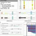
Fig. 2.1
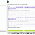
Example of a PTEN loss of expression validating a PTEN genomic loss. (a) Immunolabeling of PTEN protein (clone A2B1, Zymed® laboratories, CA, USA). The metastatic cells are PTEN negative in the presence of the positive expression of PTEN by the fibroblasts (internal positive control). (b) The SNP6.0 copy number profile shows a focal PTEN loss
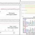
Nuclear expression of PTEN has been reported but was not considered in the frame of the SHIVA trial for PTEN expression levels’ interpretation. The cutoff to consider a case positive was at least 10 % of positive cells with a weak intensity. In the SHIVA trial, 7 cases presented a PTEN deletion and 100 a PTEN loss. One hundred three cases were available for PTEN IHC analysis. In 56 out of the 103 analyzed cases (54 %), PTEN was not expressed, signing out the genomic loss (Fig 2.2).
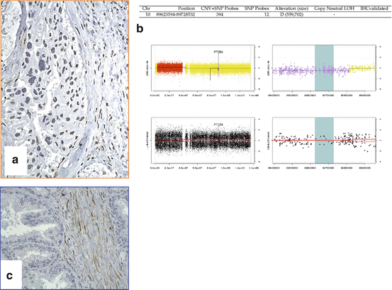

Fig. 2.2


Example of a PTEN loss of expression validating a PTEN genomic deletion. (a) Immunolabeling of PTEN protein (clone A2B1, Zymed® laboratories, CA, USA). The metastatic cells are PTEN negative in the presence of the positive expression of PTEN by the fibroblasts (internal positive control). (b) The SNP6.0 copy number profile shows a focal PTEN deletion. (c) The external positive control represented by the intracytoplasmic positivity of the normal prostatic fibroblasts
Stay updated, free articles. Join our Telegram channel

Full access? Get Clinical Tree


