Fig. 14.1
Anatomical boundaries of the central neck compartment
Within the central compartment, there are several groups of lymph nodes that are resected during a central compartment dissection. The prelaryngeal nodes are also known as the Delphian nodes and lie just anterior to the cricothyroid membrane near the pyramidal lobe of the thyroid gland. The pretracheal nodes lie anterior to the trachea but inferior to the thyroid isthmus. The paratracheal nodes are encountered posteriorly to the lateral lobes of the thyroid in close proximity to the recurrent laryngeal nerve. If these nodes are encountered more posteriorly, they are often described as retropharyngeal or paraesophageal nodes. To perform a complete CND, the prelaryngeal, pretracheal, paraesophageal and paratracheal nodes should all be resected [5].
The extent of surgical management of the central neck compartment is guided in part by preoperative investigations and clinical exam. Dissection of the central neck is commonly described by the indication for nodal resection—therapeutic or prophylactic/elective. A Level VI dissection is therapeutic if lymph node metastases are detected clinically either through preoperative imaging/exam or intraoperative findings. A Level VI dissection is considered elective if there is clinically no evidence of nodal metastases. Furthermore, CND should also be described as either unilateral or bilateral.
If any additional nodal basins are resected at the time of operation, these should also be delineated using appropriate nomenclature. Frequently, Level VII nodes are included in the resection of the central compartment for thyroid cancer. Level VII nodes are the superior mediastinal nodes that are in direct continuity with the Level VI central compartment. Level VII nodes are located between the suprasternal notch and the innominate vein [6]. Other nodal basins that may be involved by metastatic thyroid carcinoma are the lateral lymph nodes, Level II, III, IV and V. Level I, the submental nodes, are rarely involved. The lateral nodes are more readily accessible for ultrasound evaluation preoperatively than the central compartment and thus may be biopsied if they appear suspicious. This allows for confirmation of metastatic disease prior to surgical intervention, eliminating the need for prophylactic lateral node dissection. The most recent ATA guidelines state that lateral neck dissection should only be performed for patients with biopsy-proven metastatic lateral cervical lymphadenopathy (Recommendation 37, [2]).
Background: Patterns of Lymph Node Spread
The spread of thyroid cancer cells in PTC occurs mostly through lymphatics and less often via the haematogenous route. The nearest lymphatic drainage of the thyroid gland is the central compartment nodes (level VI); thus, these have the highest rate of nodal metastases for differentiated thyroid cancer [7]. Tumours located in the upper thyroid pole may spread directly to lateral compartment nodes and “skip” the central compartment. Surgeons should be aware of this pattern of metastasis when planning lymph node surgery.
A retrospective review by Machens et al. in Germany [8] saw a higher incidence of nodal metastases with increasing size of the primary tumour. In this group, 14% of patients with T1 tumours had central compartment metastatic disease, while 45% of the T4 tumours harboured central nodal metastases. The incidence of metastatic disease was increased within ipsilateral central nodes vs. contralateral central nodes (29% vs. 13%), illustrating that thyroid cancer tends to metastasize to the nodes in closest proximity.
Background: Limitations of Imaging and Clinical Exam
Ultrasound is an invaluable tool for the clinical evaluation of patients with thyroid cancer. Ultrasound helps to identify thyroid nodules with characteristics suspicious for malignancy and can be used to delineate which nodules undergo biopsy. Its utility is restricted, however, in the central compartment given anatomical constraints which limit detectability of nodal metastases. One study showed that the sensitivity of ultrasound for detection of nodal disease for papillary thyroid cancer was only 36.7% [9]. It is also more difficult during preoperative physical exam to palpate central nodal metastases given the presence of other structures anteriorly (strap muscles, thyroid gland, clavicles). For these reasons, up to 60% of patients with positive central nodes on final histological assessment histology are undetected clinically prior to operation [10].
Choi and colleagues in Korea [11] performed a retrospective analysis of 589 patients with PTC who underwent both preoperative ultrasound and CT scan to evaluate for nodal disease. All patients underwent a central node dissection at operation. After correlation with final histopathological data, they found that ultrasound had only a 47.16% sensitivity for central compartment metastases, and similarly CT had only a 41.94% sensitivity. Both modalities were much better in evaluating the lateral compartment with sensitivities near 70%. An additional finding in this study was that many of the metastatic nodes within the central compartment were small (<5 mm) thus are unlikely to be detectable on ultrasound which traditionally identifies metastases based on enlargement of the node. This finding was confirmed by Vergez and colleagues [12] who noted that in patients with positive central node compartment metastases, the largest lymph node measured 5 mm or less in 66% of cases.
It is also well known that surgeons are poor estimators of nodal disease intraoperatively. A well-cited study of surgeon detection of metastatic nodal disease during thyroidectomy for medullary thyroid cancer showed a sensitivity of only 64% [13]. It is common to have enlarged lymph nodes that are reactive to whatever concurrent pathology exists in the thyroid gland (Hashimoto’s thyroiditis, Graves disease, etc.), and these enlarged nodes may be mistaken intraoperatively as malignant. With a standard practice of routine CND on all patients, there is no need for reliance on surgeon or imaging detection of nodal metastatic disease. The most reliable method of detecting metastases is surgical resection and histopathologic evaluation.
Benefits of Elective Central Lymph Node Dissection
Decreased Local Recurrence
Performance of a therapeutic central node dissection decreases the rate of recurrence of differentiated thyroid carcinoma. The group in Dusseldorf, Germany, showed a decreased rate of recurrence when central compartment dissection was performed at initial operation for both papillary and follicular thyroid carcinomas [14]. Similarly, there is also a decreased rate of recurrence when central node dissection is performed electively. Pereira and colleagues in Barcelona reported no recurrence within the central compartment for 43 patients after either therapeutic (15/43) or elective (28/43) CND. Overall, the five patients in this study who developed recurrence (11%) all recurred laterally with no central recurrences after central node dissection at initial operation [15].
A lower rate of recurrence associated with central node dissection has been shown in multiple other studies (see Table 14.1). The group at New York-Presbyterian Hospital noted a recurrence rate of 16.7% in patients without CND vs. only 4.4% in those who underwent CND, although this did not reach statistical significance [16]. Similar to the Barcelona group, they too noted that no patients who underwent CND developed recurrence within the central compartment. Hartl and colleagues in France showed only 2% recurrence for those who underwent CND vs. 12% recurrence in thyroidectomy alone over 5 years, p < 0.001 [17]. Both cohorts in this study received similar doses of RAI and had no difference in complication rates.
Table 14.1
Comparison of recurrence rates after CND
Study | Total number of patients (n) | Recurrence without CND | Recurrence after CND | p value | CND effect on recurrence |
|---|---|---|---|---|---|
Popadich 2011 | 606 | 6.1% | 1.5% | 0.004 | REDUCED |
Barczynski 2013 | 640 | 13.1% | 4.2% | <0.001 | REDUCED |
Hartl 2013 | 246 | 12% | 2% | <0.001 | REDUCED |
Several larger studies have also examined recurrence after prophylactic CND. A multicentre study in 2011 by Popadich et al. included endocrine surgical centres in Australia, the United States and the United Kingdom [18]. This retrospective review of 606 patients with PTC >1 cm showed an overall rate of reoperation for recurrence of 5% in Group B (elective CND and total thyroidectomy) compared to 8.4% of patients in Group A, total thyroidectomy alone (p = 0.11). When analyzed as recurrence-free survival within the central compartment, there was a statistically significant improvement for those who underwent elective CND (Group B) as they did not recur within the central compartment (see Fig. 14.2). Furthermore, this study found that in the overall cohort, the performance of 20 prophylactic central neck dissections would prevent one central neck recurrence.
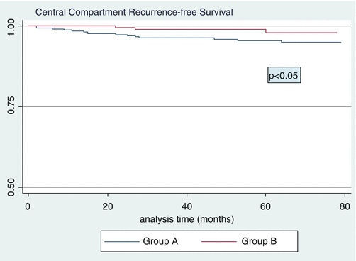
Fig. 14.2
Central Compartment Recurrence-free Survival curves for Group A (total thyroidectomy alone) vs. Group B (total thyroidectomy and CND). Group B (upper curve) had fewer reoperations within the central compartment than those in Group A (lower curve). Reprinted with permission from Popadich et al. A multicentre cohort study of total thyroidectomy and routine central lymph node dissection for cN0 papillary thyroid cancer Surgery. 2011;150:1048–57
In 2013, Barcyznski et al. published a retrospective review of 640 patients in the British Journal of Surgery [19]. Three hundred and fifty-eight of these patients underwent a prophylactic CND and were compared to 282 patients who did not undergo CND. They found that performance of a prophylactic CND was an independent predictor for improved locoregional recurrence at 10 years postoperatively, Odds Ratio 0.21. They also concluded that patients who underwent prophylactic CND had improved disease-specific survival at 98% over 10 years compared to 92.5% (p = 0.034).
Wang and colleagues in the United States conducted a large meta-analysis of 2318 patients looking at the risk of recurrence following prophylactic CND in clinically node-negative patients. While it did not reach statistical significance, there was a lower rate of recurrence in patients with CND than in those without—4.7% vs. 7.9% [20].
Decreased Postoperative Thyroglobulin Level
Thyroglobulin (Tg) level is a clinical marker of recurrent PTC that is closely monitored postoperatively by endocrinologists. An elevated Tg level after thyroidectomy and radioactive iodine ablation indicates the presence of active thyroid tissue within the body. Detectable Tg can come from either remnant thyroid tissue within the neck or from metastatic disease.
Related posts:
 Evaluation of a Thyroid Nodule
 The Rising Incidence of Thyroid Cancer: Contributions from Healthcare Practice and Biologic Risk Factors
 Molecular Diagnostic Approaches and Their Clinical Utility
 Imaging Modalities in the Diagnosis of Recurrent or Metastatic Thyroid Cancer
 Lymph Node Dissection for Differentiated Thyroid Cancer
 Pathologic Diagnosis of Thyroid Cancer
Stay updated, free articles. Join our Telegram channel

Full access? Get Clinical Tree


