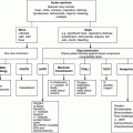
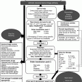
Chapter 5 Jecko Thachil Department of Haematology, Central Manchester University Hospitals NHS Foundation Trust, Manchester, UK The prothrombin time (PT) and activated partial thromboplastin time (APTT) are the commonly used screening tests for coagulation. These tests were developed to assess the coagulation system based on their classification into extrinsic (PT) or intrinsic (APTT) clotting pathways. For this reason, an abnormality in these tests would mean an abnormality in the clotting cascade, which does not always translate to clinical bleeding. At the same time, normal PT and APTT, although useful in ruling out most causes of bleeding disorders, cannot exclude some conditions including mild bleeding disorders and rare diseases like factor XIII deficiency (see Table 5.1 for an interpretation of basic coagulation tests). Table 5.1 Interpretation of clotting tests. Plt, Platelet count; N, normal; DIC, disseminated intravascular coagulation. 1 Vascular abnormalities include scurvy, Cushing’s disease and Ehlers–Danlos syndrome. 2 A mild coagulation disorder can be masked by the administration of blood products. This would include mainly mild factor VIII deficiency and some cases of von Willebrand disease. 3 PT is relatively more prolonged than APTT. Prothrombin time and APTT tests are based on restarting the coagulation process, which has been inhibited by removal of calcium, complexed to the anticoagulant citrate in the coagulation tubes. To facilitate the coagulation process, which usually occurs on phospholipid membranes (of mainly platelets) in vivo, thromboplastin is added, which forms the source of phospholipids in vitro. Time taken for the clot to form is measured in seconds and compared with normal control. This test measures the components of the extrinsic coagulation system (tissue factor; the initiator of this pathway was considered extrinsic to the body). Although it was originally devised to measure prothrombin (clotting factor II), it is now known to be affected by deficiencies of coagulation factors V, VII and X and fibrinogen, as well. The test involves adding an optimal concentration of thromboplastin (source of phospholipids) to the plasma, which has been recalcified. Thromboplastin or tissue extract used in PT was originally sourced from rabbit brain or lung, although more recently it is obtained by recombinant methods. The latter is highly sensitive to decrease in clotting factors since there is no contamination with any animal coagulation proteins. A prolonged PT in ITU is most commonly due to the lack of vitamin K. Other causes in decreasing order of frequency include: In liver disease, the PT is often the first abnormality (before APTT), since factor VII has a short half-life and its deficiency is therefore the first to be detected. In DIC, PT is more often abnormal than APTT since factor VIII levels, which can increase with conditions leading to DIC, can correct APTT. Very rarely, antibodies to factors VII, II, V and X can also prolong PT with special mention of factor V antibodies developing in those who had bovine thrombin as a haemostatic agent during surgery. Prothrombin time is generally insensitive to unfractionated heparin since the PT reagent includes heparin neutralization agents. However, if there is contamination with concentrated heparin, the PT may be prolonged. Shortened PT is almost always seen with activated recombinant factor VII treatment. Since PT is based on tissue thromboplastin reagents, depending on the tissue of origin, variability in clotting times had been noted. The use of recombinant-derived thromboplastins did not completely eliminate this variability. For this reason, the international normalized ratio (INR) was developed mainly to standardize the widespread use of the oral anticoagulant warfarin. Warfarin exerts its anticoagulant action by inhibiting the function of Vitamin K-dependent coagulation factors, which include factors II, VII, IX and X. INR uses a World Health Organization reference thromboplastin standard so that INRs measured anywhere in the world would yield similar results.
The Abnormal Clotting Profile
Introduction
PT
APTT
Plt
Condition
N
N
N
Common – normal haemostasis, vascular abnormalities1
Rare – platelet dysfunction, dysfibrinogenaemia, mild coagulation factor deficiency2
Extremely rare – factor XIII deficiency, alpha 2-antiplasmin deficiency
Long
N
N
Common – coumarin anticoagulants, vitamin K deficiency, liver disease
Rare – factor VII deficiency
N
Long
N
Common – antiphospholipid antibody, heparin, liver disease, factors VIII, IX, XI, XII deficiency, von Willebrand disease
Rare – inhibitors to the aforementioned factors, high-molecular-weight kininogen (HMWK) or pre-kallikrein (PK) deficiency
Long
Long
N
Common – vitamin K deficiency3, oral anticoagulants3
Rare – factors V, VII, X and II deficiency
Long
Long
N
Heparin, liver disease, fibrinogen deficiency
Hyperfibrinolysis
Long
Long
Low
DIC, acute liver disease
The principle of coagulation screen
Prothrombin time (PT)
INR
Stay updated, free articles. Join our Telegram channel

Full access? Get Clinical Tree


