© Springer-Verlag GmbH Germany 2018
Daniel J. Ledbetter and Paul R.V. Johnson (eds.)Endocrine Surgery in Childrenhttps://doi.org/10.1007/978-3-662-54256-9_2222. Testicular Tumors in Children
(1)
American University of Beirut Medical Center, PO Box: 11-0236, Riad El Solh, Beirut, 1107 2020, Lebanon
(2)
Department of Surgery, Division of Urology, The Hospital for Sick Children, University of Toronto, 555 University Avenue, Toronto, ON, M5G 1X8, Canada
Keywords
Testicular tumors in childrenPediatric testicular tumorsDisorders of sex developmentGonadal stromal tumorsRadical orchiectomyGerm cell tumorsThere is a growing body of evidence that shows testicular tumors in prepubertal children and infants to be distinct clinically from testicular tumors in adults, or even adolescents, where more benign pathologies are being encountered in younger patients. With testicular tumors in children being relatively rare, this observation culminated with scattered small case series, in addition to centralized tumor registries, for example, the Prepubertal Testis Tumor Registry by the Section of Urology of the American Academy of Pediatrics (1980) and national oncology trials namely, the Children’s Oncology Group in the USA, the German Society of Pediatric Oncology, and the Children’s Cancer Study Group in the UK. This has led to fine-tuning in the management of prepubertal testicular tumor by showing radical surgery to be unnecessarily aggressive. This has led in a shift in the paradigm of management in pediatric testicular tumors, where more testis-sparing surgery is being performed without compromising safety and prognosis, hence reducing morbidity of adjuvant treatment that is skipped.
Epidemiology
Testicular tumor incidence follows a bimodal age distribution, peaking at around two years of life then during early adulthood [1]. Prepubertal testicular tumors account for 1–2% of pediatric solid tumors with an incidence of 0.5–2.0 per 100,000 children [2]. Mortality rates from pediatric testicular tumors are low, with one death per ten million per year. Prepubertal testicular cancer survival is about 99% at 5 years [3]. When looking into individual frequencies of pathologies, one has to be aware of inherent selection bias in underreporting benign cases to tumor registries leading to discordant reporting. Indeed, while earlier registry data showed more frequent malignant pathologies: 60% yolk sac tumors versus 40% benign and 25% teratoma [4], Pohl et al. surveyed four major pediatric centers in North America and found higher frequency of benign pathologies in unselected testicular lesions up to 75%, namely, 50% teratoma, 15% epidermoid cyst, 5% juvenile granulose cell tumors with less than 5% each of Leydig cell tumors, Sertoli cell tumors, and mixed gonadal stromal tumors. Malignant yolk sac tumors in this survey were only 15% [5].
Risk Factors and Associations
The exact causes for testicular cancers are not known. Few risk factors are identified that contribute to an increased incidence of testicular cancer while other associations are under study with no solid evidence in literature.
Cryptorchidism
The United States National Cancer Institute lists cryptorchidism, or undescended testicle, as a main risk factor for testicular cancer. Statistically, 5% of testicular cancers are associated with a history of cryptorchidism [6]. A cryptorchid testicle may be genetically and anatomically predisposed to abnormal structure and function, leading to testicular dysgenesis syndrome (TDS), which increases the risk for malignant degeneration, as well as poor semen quality and fertility implications [7]. In routine practice, most pediatric urologists would strive to perform orchidopexy prior to age 2, and would counsel families whose kids present with cryptorchidism about the risk of malignancy. Wood and Elder [8] found that the relative risk of testicular cancer in cryptorchidism is 2.75–8. Patients who undergo orchiopexy by ages 10–12 years have a relative risk for testicular cancer of 2–3, while patients who undergo orchiopexy after age 12 years or no orchiopexy are 2–6 times as likely to have testicular cancer as those who undergo prepubertal orchiopexy. A normally descended testis with contralateral cryptorchidism has no increased risk of testis cancer. Persistent inguinal or abdominal testes are at higher risk for seminoma (74%), while corrected cryptorchid or scrotal testicles that undergo malignant transformation are most likely to become nonseminomatous (63%).
Disorders of Sexual Differentiation
Patients with disorders of sex development (DSD), previously referred to as intersex disorders, are a unique subpopulation that is at an increased risk of gonadal tumors. DSD has been noted to be a main risk factor for type II germ cell tumors (GCT), namely, seminoma/dysgerminoma and non-seminoma (e.g., embryonal carcinoma, yolk sac tumor, choriocarcinoma and teratoma [9]. DSD male patients often have chromosomal abnormality that predisposes to genital ambiguity, hypovirilization, and a spectrum of gonadal dysgenesis. The prevalence of GCT is 15% in partial androgen insensitivity, but more than 30% in gonadal dysgenesis. Complete androgen insensitivity and ovotesticular DSD patients develop malignancies in 0.8 and 2.6%, respectively [10]. Recently, undifferentiated gonadal tissue has been implicated to be a precursor of gonadoblastoma. This transformation requires a defined region on the Y chromosome (the GBY region) which included the gene for testis-specific protein on the Y chromosome (TSPY) which is evolving as a marker for gonadoblastoma and GCT [11]. Aberrant TSPY expression increases protein synthesis, stimulates cell proliferation, and promotes tumorigenicity by binding to type B cyclins, enhancing an activated cyclin B-CDK1 kinase activity, resulting in a rapid G [2]/M transition in the cell cycle. Abnormal expression of TSPY gene in a DSD testis disrupts the normal cell cycle regulation leading to tumorigenesis [12].
Environmental Factors
Environmental factors are always connected to cancer formation, although in many cases the level of evidence is not compelling, with the data being largely mixed and inconclusive and the real accusable factors remaining elusive. Caucasians are more noted to develop testicular cancer than other ethnic groups. Additionally, there is a remarkable worldwide variation of testicular cancer incidence according to geographical region. For instance, while Denmark has among the highest incidence of testicular cancer in the world, the risk of testicular cancer was significantly lower among first generation immigrants to Denmark, versus men born in Denmark to immigrant parents and Danish men with Danish parents [13]. The higher incidence of testicular cancer in the western world can be partly attributed to the rise in industrial chemicals over the last century. Prenatal exposure (or even during adolecense or adulthood) to such chemicals, namely, polychlorinated biphenyls, DDT, phthalates, hexachlorobenzene and other fat soluble organochlorines, may predispose to testicular cancer, as they were found in higher concentrations in mothers of males with testicular cancer [14]. A hypothesis, to explain why chemicals would influence testicular cancer development, is that they interfere with or mimic hormonal signaling pathways in utero or later, leading to aberrant development or differentiation, hence they are collectively termed, endocrine disrupting chemicals (EDC) [15]. Further studies have noted increased association with testicular cancer in obesity [16]. Maternal consumption of alcohol during pregnancy, but not smoking or coffee intake, was associated with higher risk of testicular cancer in their sons [17]. Recently, an association between marijuana use and GCT, particularly non-seminoma, was noted [18].
Microlithiasis
With the increased utility of ultrasonography in clinical medicine, the concern about testicular microlithiasis (TM) association with testicular cancer rose as microlithiasis is often an incidental finding affecting up to 14% of asymptomatic adult men and up to 2% of asymptomatic boys [19, 20]. Whenever this entity is diagnosed, it compels the clinician to provide follow-up, counseling and prognosis, namely because TM has been associated with certain genital pathologies: cryptorchidism in 10% [21], infertility in up to 23% [22], McCune–Albright syndrome in 60% [23], and testicular tumors in an alarming 45% [24]. TM is defined as foci of calcification within the seminiferous tubules that may represent tiny foci of dysgenetic tissue that could potentially progress into carcinoma in situ. Although the association of TM with tumor formation is mainly demonstrated in adults, only four scattered case reports were cited in children [25–28], making the association inconclusive. It is worth noting that testicular tumors have never been identified during prospective follow-up of a cohort followed up for TM, in either adults or children. Although some investigators found long-term follow-up of children with TM to be anxiety-provoking, not cost effective and with low yield [29], others still recommend ultrasound surveillance, physical examination and repeated patient education, with no well-defined protocols.
Clinical Presentation and Evaluation
History and Physical Examination
The most common presentation of testicular tumor is a painless testicular mass, noted in 88% in the Testis Tumor Registry. The parents maybe instrumental in detecting the mass, or it may be detected on a routine physical examination by the primary health care provider. Around 10–15% of patients would present with a coincidental or tumor-related hydrocele at presentation. A hydrocele may delay the diagnosis of a testis tumor. It is crucial, thus, to obtain an ultrasound in boys with a hydrocele precluding reliable palpation of the testicle. Pain is uncommon and may be due to acute bleed into the tumor or a rapid growth.
Physical examination is integral to the evaluation of a testicular mass and is performed with a broad differential diagnosis in mind to rule out a hernia, hydrocele, inflammatory/infectious orchitis (e.g., mumps), as well as the surgical emergency of acute testicular torsion. Physical examination reveals a hard mass in the testicular parenchyma. However, small masses deep in the parenchyma may be difficult to palpate, and would require sonographic assessment. Testicular masses must be differentiated from benign extratesticular lesions, whether benign (e.g., epididymal cyst) or malignant (e.g., rhabdomyosarcoma). The genitalia and other body parts should be carefully examined for signs of androgenization or feminization. Although metastatic disease at presentation is uncommon, examining groins, axillae and neck for lymphadenopathy, ausculting the lungs for abnormal sounds, eliciting bony pain and even a brief neurologic exam may yield positive findings in the rare occasion of metastasis and is considered good practice. Symptoms or signs of involvement at these anatomic locations guide subsequent radiographic evaluation.
Imaging and Other Diagnostics
An initial imaging with ultrasound remains the cornerstone evaluation of a testicular mass with a detection rate approaching 100%. High frequency window of 7.5–10 MHz can be beneficial in elucidating particular sonographic features of testicular versus paratesticular neoplasms [30] and while sonographic description of specific testicular tumors is available in radiologic literature, findings are too inconsistent to allow for a definitive diagnosis. Benign tumors are well demarcated with sharp borders and decreased Doppler signal. Epidermoid cysts have an echogenic rim with mixed echogenic or hypoechoic center [31]. Yolk sac tumors have homogeneous hypoechoic solid appearance, but may have other sonographic appearance. Ultrasound would also distinguish testicular tumors from extra-testicular lesions. The extent of testicular involvement can also be appreciated, a criteria that would be considered among others, if a testis-sparing surgery is contemplated.
Tumor markers are vital diagnostic clues in evaluating testicular tumors. Alpha feto-protein (AFP) is the most important marker in evaluating and following-up prepubertal testicular tumors. It is precursor protein of albumin, synthesized by the yolk sac and fetal liver and gut. AFP has a half- life of 5 days and is specific for yolk sac tumors, the main malignant tumor in this age group. Levels are elevated in 80–90% of children with a yolk sac tumor, and its preoperative elevation precludes testis-sparing surgery. The utility of AFP in infancy, however, can be confusing since an elevated level in a boy less than 1 year of age does not rule out the possibility of a benign tumor, like teratoma. AFP’s known half-life of 5 days is attained around 4 months of life and is longer before that. Serum AFP would be normally as high as 50,000 ng/mL in neonates, 10,000 ng/mL by 2 weeks of age, and 300 ng/mL by age of 2 months [32]. While yolk sac tumor and teratoma elevate AFP, data from the Testis Tumor Registry did not show any teratoma diagnosed after 6 months of life to elevate AFP more than 100 ng/mL [4]. This observation highlights the decision-making to remove or spare the testis.
The other classical marker in testicular tumors is the beta subunit of the human chorionic gonadotropin (hCG) produced by the syncytiotrophoblast of the placenta. While this is useful in postpubertal and adult testicular tumors, it is not very useful in children, particularly because choriocarcinoma and embryonal carcinoma, both secreting hCG, are rare [33].
Because the majority of testicular tumors in prepubertal boys are, by and large, benign, the evolving strategy of evaluation is to defer metastatic workup until a tissue diagnosis is obtained at time of orchiectomy. This would spare the patient the unnecessary exposure to a CT scan if his pathology turns benign. Preoperative CT scan or MRI is warranted, however, if AFP is elevated, raising the suspicion of a yolk sac tumor. Because lung metastasis is present in 20% of yolk sac tumors, chest imaging is warranted.
Surgical Approach
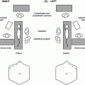
Regardless of the eventual surgical management of testicular tumor by radical orchiectomy or testis-sparing surgery (partial orchiectomy or enucleation of tumor), the initial surgical approach and exposure is the same [34]. An ipsilateral inguinal incision is made along a Langer’s line crease of minimal tension. The incisional length can be extended at surgeon’s judgment to improve exposure, particularly to deliver large tumors. The incision is deepened using electrocautery through Scarpa’s fascia to reach the external oblique aponeurosis. In younger patients, Scarpa’s fascia is well developed and may be mistaken for the external oblique fasica. The external oblique fascia is sharply incised along its fibers to open the external inguinal ring, paying attention to preserve the underlying ilioinguinal nerve which provides cutaneous sensation to the inner aspect of the upper thigh. The spermatic cord is identified and bluntly dissected inferiorly to expose the pubic tubercle. Incising the cremasteric sheath longitudinally allows for easy isolation of the cord and precludes the need to repair the internal oblique which constitutes the canal floor and gives rise to the cremasteric fibers. The proximal spermatic cord is controlled with noncrushing clamp or a vessel loop/Penrose drain wrapped around it as a tourniquet. Theoretically, this prevents dissemination of potential malignant cells into bloodstream from testis manipulation, although little evidence supports this “ritual” practice. The scrotal neck is stretched and Scarpa’s fascia incision may be extended while the testis is delivered out of the incision by gently pushing it out of the scrotum. This step may be challenging in large tumors. The gubernaculum is divided with electrocautry while carefully maintaining an intact tunica vaginalis covering the testis. At this point and according to the clinical scenario, the procedure can proceed as radical orchiectomy or a testis-sparing procedure.
Radical Orchiectomy
In the clinical scenario of a malignant tumor, almost invariably a yolk sac tumor evidenced by preoperative elevation of AFP, the above procedure is completed as a radical orchiectomy. The spermatic cord is doubly clamped, divided and the vas and vessels are doubly ligated above the internal inguinal ring level, using 2-0 or 3-0 nonabsorbable sutures cut sufficiently long. This allows identification and excision of the cord intraperitoneally, if later retroperitoneal lymph node dissection is undertaken. The testis and cord are sent in formalin for definitive pathological analysis. The abdominal defect is closed using absorbable sutures: the conjoined tendon may be reinforced by suturing it to the shelving edge of the inguinal ligament then the external oblique incision edges are closed together or imbricated. Scarpa’s fascia is approximated and skin is closed in a running subcuticular fashion using absorbable sutures. No drain is necessary. Testicular prosthesis insertion in children is deferred until adolescence. Matching size prosthesis is offered in late adolescence for willing patients, old enough to consent and after near maximal compensatory growth of the contralateral testicle has occured.
Testis-Sparing Surgery
With one-third to up to 40% of prepubertal testicular tumors being benign, a growing trend is emphasizing testis-sparing surgery. Any prepubertal boy with age-adjusted normal AFP is a candidate of testis-sparing surgery. Other indications include small tumors less than 2.5 mm in diameter, a tumor in a solitary testis, bilateral malignant germ cell tumors or a non-germ cell tumor, regardless of size or laterality [35]. It has been observed that after partial orchiectomy for large benign tumors, a seemingly insignificant rim of normal tissue tends to get voluminous with follow-up, after the tumor is removed [36, 37].
For testis-sparing procedure, a cautious meticulous approach to potential malignancy is maintained. With the cord controlled, the testis is further draped to isolate it from the rest of the surgical field. The tunica vaginalis is opened and the testis is gently palpated for the tumor. Intraoperative ultrasound is useful to localize small tumors that are not convincingly palpable. A tunica albugenia incision is strategically done with knife in a way to allow access to the tumor with the least disruption of normal parenchyma. The tumor is enucleated or wedge-resected as feasible and sent for frozen section analysis. If the pathology is benign, the tourniquet is released and hemostasis of base of resection is ensured by suture ligatures and minimal bipolar cautery. The tunica albugenia is closed with fine running absorbable sutures. The testis is returned to scrotum in proper orientation and abdomen is closed. If frozen section analysis is suspicious for malignancy or is not conclusive, radical orchiectomy is completed as described above.
Staging
When pathology confirms a malignant tumor, further evaluation and adjuvant therapy may be needed. Therefore, staging of malignant tumors emerges as a method to categorize patients into treatment groups and to predict their prognosis. Although a classical TNM (tumor, nodes and metastasis) staging does exist for testicular cancer, it is infrequently used, owing to the declining utilization of radical retroperitoneal lymph node dissection (RRPLND) in pediatric patients because of evidence of limited added therapeutic benefit and significant potential long-term morbidity. However, other staging systems have been proposed, including a panel system of the Children’s Oncology Group and Pediatric Cancer Group [38]. This staging system of testicular, as well as paratesticular tumors, employs pathology findings, imaging, and serial evaluation of tumor markers. Stage 1 includes local disease and normalization of markers after complete resection. Stage 2 is assigned for a trans-scrotal orchiectomy, microscopic disease in scrotum or cord less than 5 cm from proximal resection end, less than 2 cm retroperitoneal lymph node and/or persistent elevation of markers. Stage 3 includes retroperitoneal lymph node(s) larger than 2 cm in diameter and stage 4 represents distant metastasis. Of note, 80% of children with yolk sac tumor have stage 1 disease.
Types
Testicular tumors may be classified according to their histologic origin and their malignant potential.
Germ Cell Tumors
Teratoma
Teratoma is the most common benign prepubertal testicular tumor presenting at a median age of 1 year with a range of birth to 18 months. Teratomas consist of the combination of the three embryological germ-cell layers: ectoderm, mesoderm, and endoderm, giving rise to epithelium, cartilage, fat, bone, muscle, and neural elements, thus appearing heterogeneous sonographically. Cysts and calcifications on ultrasound are suggestive but are neither sensitive nor specific. Microscopically, prepubertal teratomas are predominantly mature, but immature teratomas (with embryonal or incompletely differentiated tissue, e.g., primitive neuroectoderm) have been reported [39].
Prepubertal teratoma is clinically and histologically distinct from its adult counterpart and is considered to have an almost universal benign behavior. While clinical metastasis is reported in 60% of adult teratoma [40, 41], only scattered case reports describe metastasis in prepubertal teratoma. Histologically, intra-tubular germ cell neoplasia (ITGCN) or carcinoma in situ is not found in seminiferous tubules of pediatric testes harboring teratoma, unlike the findings in adult testes with teratoma where 88% have ITGCN [42]. Even at a molecular level, adult teratoma shows complex cytogenetic aberrations that are not observed when prepubertal teratomas are subjected to conventional chromosomal or array-based hybridization studies [43], thus demonstrating the benign behavior of prepubertal teratoma. As such, testis-sparing surgery with excisional biopsy of teratoma is the only management required in prepubertal patients. However, in peripubertal boys with histologic evidence of pubertal changes, an orchiectomy should be performed because teratomas are potentially malignant in that age group, similar to adults. It is worth mentioning that malignant behavior of teratoma reported in literature, albeit rare, occurred with immature teratoma: one case of metastasis in a 3 month old boy with immature teratoma in a cryptorchid intra-abdominal testis [44], and 2 cases of somatic malignant neuroectodermal associated with testicular immature teratoma in a 20 month and 12 year boys [39].
Epidermoid Cysts
Epidermoid cysts may be related to well-differentiated teratomas and are benign regardless of age. They account for 3% of primary testicular tumors. They are of ectodermal origin, unlike teratomas that exhibit all three germ cell layers. On ultrasound, they appear as discrete intratesticular cystic masses with heterogeneous echogenic debris secondary to keratinized squamous epithelial deposits, Infrequently, they appear as homogeneous masses. Again, ultrasound findings are suggestive but never diagnostic. Testis-sparing surgery is sufficient, and epidermoid cysts are easily enucleated from surrounding testicular tissue [45], that never shows ITGCN [42]. Once pathology is confirmed, surveillance is not needed.
Yolk Sac Tumors
Yolk sac tumor is clinically important as it accounts for nearly all malignant prepubertal testicular tumors. The name yolk sac tumor has replaced numerous previous names that were confusing: clear-cell adenocarcinoma, extraembryonal mesoblastoma, mesoblastoma vitellinum, endodermal sinus tumor, juvenile embryonal carcinoma, orchioblastoma, and archenteronoma. Histologically, yolk sac tumors are yellow-gray, friable, predominantly solid tumors with a characteristic but highly variable range of histological patterns. Characteristic findings include focal areas of positive AFP immunostaining, prominent intracellular and extracellular hyaline globules, periodic acid-Schiff positive globules and Schiller–Duval bodies which are comprised of small central blood vessels surrounded by two layers of tumor cells [46].
Yolk sac tumors in prepubertal patients behave distinctly from their postpubertal counterparts. In children, they are predominantly pure in histology unlike in adults where they are mixed with other histological types. The majority of prepubertal tumors (85%) are stage 1 at presentation versus only 35% of postpubertal tumors [47]. Metastasis of yolk sac tumors follows a hematogenous spread, as 20% of prepubertal patients have lung metastases versus 4–6% of adults. While one-third of children with metastatic disease have it confined to the retroperitoneum, 50% have hematogenous spread without retroperitoneal involvement. As a result RRPLND, a critical adjuvant surgery in metastatic disease in adults, not as widely used in the management of metastatic disease in children [48]. Even at a molecular level, prepubertal yolk sac tumor behaves differently than its postpubertal counterpart. A chromosomal gain in the short arm of chromosome 12p has been noted in nearly all malignant adult germ cell tumors, a finding that is not demonstrated in prepubertal yolk sac tumor, yet it is characterized by other distinct changes on chromosomes 1, 6, and 20 [43].
Gonadal Stromal Tumors
Given the rarity of gonadal stromal tumors in children, as well as in adults, no rigorous guidelines are available to aid management, except for some case reports and small case series. The various types of stromal tumors are generally benign, though some tumors, particularly sertoli cell tumor, were shown to exhibit malignant behavior.
Related posts:


Stay updated, free articles. Join our Telegram channel

Full access? Get Clinical Tree


