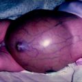
Fig. 21.1
Intra-vaginal torsion
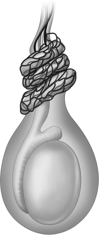
Fig. 21.2
Extra-vaginal torsion
The intra-vaginal pattern has been traditionally ascribed to a predisposing anatomical variant termed the “bellclapper” deformity, shown in Fig. 21.3a, b. However, whilst the concept of “horizontal lie” and the “bell clapper testis” are long established in conventional teaching, it is important to appreciate that the distinction between “horizontal” and “normal” orientation of the testis is inherently subjective. The reproducibility of this finding and the existence of “horizontal lie” as a distinctive anatomical variant have never been objectively validated in studies of patients and age-matched normal controls. The bell clapper deformity has, however, been the subject of a formal autopsy study reported by Caesar and Kaplan [1]. The relevant anatomical features of 101 testes were documented in series of 51 consecutive postmortem examinations in subjects ranging in age from 1 day to 75 years at the time of death. On the basis of the anatomical relationship between the testis and attachment of the tunica vaginalis these authors found that 12% of all testes demonstrated the “bell clapper” deformity. They concluded that whilst this subset of the population may be at risk for torsion “other factors in addition to anatomic predisposition must be involved”. Similar findings were reported by Ishizuka et al. [2]. In males with normally descended testes the “bell clapper” deformity almost certainly represents one end of the spectrum of normal anatomical development rather than a distinct pathological entity. It is unclear why a small percentage of such individuals experience torsion, whilst the majority of do not.
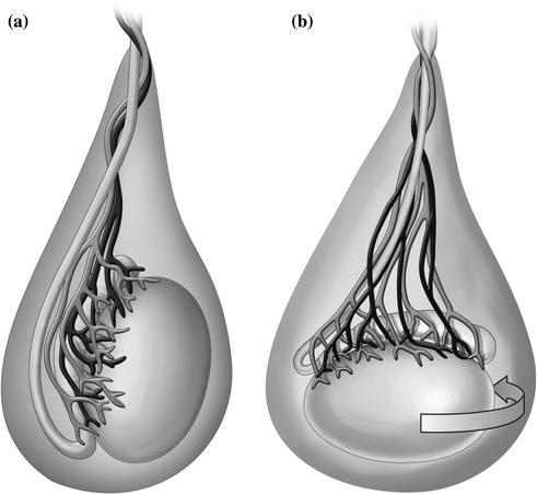

Fig. 21.3
a Normal lie. b The bellclapper deformity
Genetic Factors
The familial occurrence of testicular torsion is well documented in the literature, with particular emphasis on siblings (notably identical twins) [3, 4]. It is seems likely that this does reflect agenuine genetic basis in affected families since the incidence is greater than might be expected by chance. The mechanism(s) whereby the expression of genetic factors lead to an increased predisposition to torsion is entirely unknown.
Precipitating Factors
A number of precipitating factors have been implicated, including trauma, recent exercise and cold weather. In the series reported by Williamson [5], a history of trauma (e.g. sporting injury) and recent exercise was noted in 8 and 11% of cases, respectively. However, it is difficult to assess their significance as precipitating factors since exercise and sporting injury are relatively common events in the lives of adolescents and young adults. In the majority of instances no obvious precipitating factor can be identified and Williamson observed that “sometimes, the onset of symptoms occurred during sleep.”
Incidence
In the absence of reliable population-based data it is very difficult to derive an accurate figure for the true incidence of testicular torsion. From their analysis of 670 patients treated in Bristol, United Kingdom, between 1960 and 1984 Anderson and Williamson [6] calculated the annual incidence in their local population as 1 per 3942 males under the age of 25 years. The incidence figure of 1:4000 has been widely cited in subsequent publications.
Data collected prospectively by the Department of Health for England over an 11-year period from 1994 to 2005 indicated that the number of hospital admissions in England for “surgery for testicular torsion” in patients aged 0–17 years remained broadly constant over this period averaging 1010 hospital admissions per annum (range 884–1127) [7]. An approximation based on these Department of Health data and the annual birth rate in England yields an estimated incidence which is broadly consistent with Anderson and Williamson’s figure, i.e. 1:3000 to 1:4000. With regard to neonatal torsion an incidence of 6:100,000 (1:17,000) was reported by John et al. [8] who analyzed data from the Mersey region of the United Kingdom collected over a 13-year period. The very limited published evidence does not identify any obvious geographical variation or difference between racial groups.
Testicular torsion is characterized by a bi-modal age distribution. A small peak is observed in the newborn period but the condition occurs most commonly during adolescence, with the peak incidence around 15 years of age. In the age range 13–21 years testicular torsion has been reported to account for at least 90% of all cases of acutely presenting scrotal pathology [9]. By contrast, the differential diagnosis in pre-pubertal boys is more varied, with torsion of the Hydatid of Morgagni accounting for approximately 50% of cases. Nevertheless, torsion of the testis is also an important cause of acute scrotal pathology throughout childhood constituting 34% of cases of acute scrotal pathology in a series of boys aged 0–12 years reported by Nour et al. [10].
Pathophysiology
Our understanding of the events occurring at a vascular and cellular level is largely derived from experimentally induced torsion in rat, porcine and canine animal models [11–15]. These studies have demonstrated that 720° torsion reliably produces complete cessation of perfusion, whereas the degree of ischemia produced by less severe degrees of experimentally induced torsion is more variable.
There appears to be some degree of inter-species variation in the timescale of ischemic tissue damage but whether this reflects differences in vascular anatomy or genuine differences in susceptibility of seminiferous tissue to ischemia is unclear. Nevertheless, it seems reasonable to assume that the consequences of 720° torsion observed in pigs and dogs can be reliably extrapolated to man.
Mechanisms of Tissue Damage
Ischemia
Spermatogenesis is an extremely active process capable of generating approximately 100–200 million sperm per day [16]. This high level of metabolic activity is mirrored in correspondingly high rates of oxygen consumption by the germinal epithelium and in its marked vulnerability to periods of ischemia. The tissues comprising the testis exhibit preferential sensitivity to ischemia with the germ cell population being the first to be lost, followed by the Sertoli cells and the Leydig cells [15]. Because the germ cells and Sertoli cells comprise most of the volume of the normal testis the extent of any reduction in testicular volume can be regarded as a sensitive indicator of severity of ischemic tissue damage. The testis receives its blood supply by long, small calibre artery with a relatively high vascular resistance. As a consequence, capillary pressure in the testis is only marginally higher than the venous pressure and oxygen tension within the testis is low, particularly in relation to the high metabolic demands of spermatogenesis within the adult testis. Whilst this may be beneficial in conferring protection against oxygen-radical mediated damage to sperm DNA under normal perfusion it may also explain why testicular tissue is so susceptible to relatively short periods of ischemia.
Bergh et al. [12] found that even a 30% reduction in perfusion was sufficient to induce cell death of spermatogonia and early primary spermatocytes in the adult rat testis. Heindel et al. [13] documented reductions in sperm production and testicular weight in rats following 720° torsion maintained for as little as one hour—despite subsequent restoration of perfusion to levels comparable to those prior to the induction of torsion. In addition to its duration, the severity of torsion appears to be an important determinant of outcome. Heindel et al. [13] found that experimentally induced unilateral torsion of 720° or more had a significant impact on subsequent fertility, whereas no effect was observed following torsion of 360°.
Reperfusion Injury
In addition to tissue damage mediated by ischemia, further damage occurs as a consequence of reperfusion injury following de-torsion and restoration of blood supply to the testis. Reactive oxygen species, such as H2O2, OH, and O2, and reactive nitrogen species (RNS) such as NO and ONOO have a well-recognized potential to cause cellular injury and one of the key enzymes involved in early reperfusion injury is thought to xanthine oxidase [17–19].
The administration of exogenous antioxidants, including selenium, l-carnitine and others has been shown to reduce the severity of testicular damage in experimentally induced torsion in animal models [20–25]. Inhibition of xanthine oxidase by allopurinol has also been shown to exert a protective effect [26, 27]. To date, the use of these and other agents to ameliorate testicular damage has been limited to experimental models but it is possible that anti oxidant therapy might find a role in clinical practice in the future—particularly when the testis is left in situ following de-torsion.
The Contralateral Testis
A history of unilateral torsion is associated with an increased probability of impaired spermatogenesis in the “healthy” contralateral testis.
Although this phenomenon has been studied both clinically and experimentally, the possible mechanisms remain unclear and published findings have sometimes been inconsistent or frankly contradictory. Nevertheless, there is a sufficient evidence to pose the theoretical question of whether it might be more beneficial for fertility to remove a potentially salvageable testis (to protect spermatogenic function in the contralateral testis) rather than leave it in situ?
A detailed analysis of evidence is beyond the scope of this chapter but the possible mechanisms of impaired spermatogenesis can be summarized as follows.
Reduced Perfusion
On the basis of a number of experimental studies [28–32], it has been postulated that changes in perfusion which have been observed in the contralateral testis occur in response to toxic vasoactive agents released into the circulation from the affected testis. This occurs particularly at the time of de-torsion when the arterial perfusion and venous drainage are restored to ischemic testicular tissue.
AutoImmune Damage
Seminiferous tissue is immunologically privileged and any breach of the blood-testis barrier incurs the risk of antigens being exposed to the immune system—thus invoking an autoimmune response. This phenomenon has been demonstrated following experimental torsion in rats [33] and, in addition it has been shown that immune mediated damage to the contralateral testis can be ameliorated by administration of immunosuppressive agents, including Dexamethasone and Cyclosporine [24, 25]. The clinical significance (if any) of auto-antibodies in man is unclear. Mastrogiacomo et al. [34] reported the presence of circulating agglutinating antibodies in 20% of 25 patients evaluated 6 months to 7 years after torsion but found no correlation between the presence of these antibodies and sterility. The reported incidence of detectable antisperm antibodies varies considerably in different series and some authors have been unable to identify auto-antibodies in any of their patients despite using standardized laboratory techniques [35]. In a study of the late outcome of pre-pubertal testicular torsion, Puri et al. reported that, of patients who were married, all had fathered one or more children. In unmarried patients, semen analysis was normal in 10 out of 13. Anti sperm antibodies were not found in any of their patients. These authors concluded that pre-pubertal torsion does not cause autosensitization or diminished fertility [36].
Congenital Dysplasia
Biopsy findings have suggested that the impairment in semen quality in men with a history of unilateral testicular torsion might be explained by pre-existing congenital dysplasia in the contralateral “healthy” testis. Hagen and associates reported abnormal histological appearances in 30 out of 34 biopsies of contralateral testes obtained at the time of surgery for torsion [37]. Likewise, Anderson and Williams [38] found pre-existing histological damage in 20 out of 35 patients, prompting them to conclude that “testes prone to torsion already show impaired spermatogenesis”.
Reduced Germ Cell Mass
Can the reduction in semen quality observed in men who have undergone orchidectomy for torsion simply be explained by a quantitative reduction in seminiferous tissue? Ferreira and associates assessed the semen quality of 54 men aged between 19 and 42 years who had previously undergone unilateral orchidectomy for a variety of different indications including torsion. A significant and comparable reduction in sperm concentration was observed in all groups regardless of the indication for unilateral orchidectomy [39].
In summary, whilst follow-up studies in men with a history of torsion have consistently demonstrated evidence of impaired semen quality the causative mechanism(s) remain unclear.
Clinical Presentation
Torsion characteristically presents with severe pain of sudden onset accompanied by tenderness and swelling of the testis. The pain is often referred to the ipsilateral inguinal region and/or lower abdominal quadrant thus creating the potential for an erroneous diagnosis of appendicitis when right sided-or gastroenteritis in the case of left-sided torsion. This confusion may be compounded by a history of vomiting, which is feature of around 40% of cases of torsion. Examination of the testes should therefore be included as a mandatory part of the clinical examination of any child or adolescent presenting with sudden onset inguinal or lower abdominal pain.
It is important to note, however, that less than half of patients present with the classic “full house” of symptom and physical signs. Confusingly, pain may sometimes be minimal or absent in the early stages of torsion—particularly in younger children.
The key clinical findings consist of acute tenderness and swelling of the affected testis—which may be lying in an elevated position within the scrotum as a consequence of cremasteric contraction and physical shortening of the twisted cord. Inflammatory changes in the scrotal skin then ensue, in response to ischemia and the onset of necrosis within the underlying testis.
Differential Diagnosis
Testicular torsion accounts for around 90% of all cases of acute scrotal pathology in adolescents [9] and for this reason surgical exploration should be undertaken urgently unless there is convincing evidence of an alternative diagnosis. By contrast, the differential diagnosis is wide ranging in the pre-pubertal age group and encompasses the following;
Torsion of Hydatid of Morgagni (Torsion of a Testicular Appendage)
This typically presents with pain that is less severe, more localized and more insidious in onset than torsion of the testis. In a minority of cases, hemorrhagic infarction of the Hydatid of Morgagni is associated with visible discolouration—the so-called “blue dot” or “blue pea” sign. Although pathognomonic, this sign is more frequently absent or masked by edema and erythema of the overlying scrotal skin. An experienced clinician can often diagnose the condition on clinical grounds (with confirmation by ultrasonography if available). It is important to stress, however, that if the diagnosis cannot be established with certainty, urgent surgical exploration is indicated to exclude testicular torsion. Surgery may also be considered if the symptoms are slow to resolve on conservative management.
Epididymo-Orchitis
This diagnosis should be viewed with caution unless there is strong supportive evidence such as clear evidence of frank urinary infection and/or history of an underlying abnormality such as persistent Mullerian remnant or neuropathic bladder. Epididymo-orchitis is accompanied by marked tenderness, testicular tenderness and scrotal erythema. Other features may include fever, urinary symptoms and systemic ill health. Examination of the urine usually identifies evidence of infection. By contrast to testicular torsion the appearances on Doppler ultrasound are characterized by hyperemia rather than absent perfusion.
Idiopathic Scrotal Edema
This distinctive condition of unknown aetiology is virtually confined to young children. In contrast to the other causes of scrotal swelling and erythema in this age group, idiopathic scrotal edema typically gives rise to remarkably little pain or distress.
For the experienced clinician, the diagnosis rarely causes difficulty but where appropriate the use of scrotal ultrasonography will reliably distinguish idiopathic scrotal edema from other forms of acute scrotal pathology.
Other Causes
Other causes of acute scrotal pathology in the pre-pubertal age range include incarcerated hernia, acute hydrocele, Henoch–Schonlein vasculitis, and mumps orchitis. In adolescents the main differential diagnosis at the younger end of this age range is torsion of the hydatid of Morgagni, whereas epididymo-orchitis (possibly in conjunction with sexually transmitted disease) should be considered in the mid to later teenage years.
Investigation
The principle “investigation” is urgent surgical exploration—particularly in the post pubertal age group (in whom testicular torsion accounts for more than 90% of cases of acute scrotal pathology). Diagnostic imaging may, however, play a role in pre-pubertal boys if the clinical picture points strongly to an alternative diagnosis (e.g. torsion of the hydatid of Morgagni, epididymo-orchitis). However, if there is any suspicion of torsion, urgent exploration is still indicated in this age group.
Doppler ultrasonography is the modality of choice, but this investigation is operator-dependent and it may be difficult, if not impossible, to obtain urgently on an “out of hours basis”. Surgical exploration should never be delayed to facilitate ultrasonography if the clinical evidence raises the possibility of testicular torsion. Isotope imaging using intravenous Pertechnetate, labelled with Technetium-99M is of historical interest only and plays no useful role in current practice.
Management
Attempted external manual de-torsion is neither desirable nor feasible in children or adolescents. Time is of the essence and appropriate anesthetic precautions should be taken rather than defer induction of anesthesia because of recent oral intake. Following delivery of the testis (via a midline or scrotal compartment incision) and de-torsion, a decision must be taken on whether to conserve or remove the testis. Relevant factors include; duration of the clinical history, appearance of the testis and presence or absence of arterial bleeding on incision of the tunica albuginea. Some authors have used intra-operative Doppler ultrasonography [40–42], but this does not appear to offer any significant advantage over clinical criteria in predicting potential viability. However, one study found a strong correlation between testicular parenchymal homogeneity or heterogeneity and viability or non-viability, respectively [42].
The available evidence suggests that where doubt exists it is preferable to err on the side of orchidectomy. If the decision is taken to conserve the testis, fixation of the testis to the scrotal wall and the septum is performed using non-absorbable sutures at three sites (three point fixation). A similar technique is employed for prophylactic fixation of the contralateral testis (to obviate the risk of asynchronous contralateral torsion).
Stay updated, free articles. Join our Telegram channel

Full access? Get Clinical Tree


