Fig. 12.1
Structure of morphine. “Morphin – Morphine” by NEUROtiker – Own work (Licensed under Public Domain via Commons – https://commons.wikimedia.org/wiki/File:Morphin_-_Morphine.svg#/media/File:Morphin_-_Morphine.svg)
12.2.2 Oxycodone
Oxycodone is a phenanthrene class opioid. Its structure is shown in Fig. 12.2. It is marketed as oxycodone alone or combined with acetaminophen, ibuprofen, or aspirin. The prudent approach is to give the oxycodone alone and give an adjuvant medication separately. This allows titration of the oxycodone without the risk of unnecessary toxicity from increasing dosage of the adjuvant drug. The receptor site of activity of oxycodone is controversial. Animal studies suggest it primarily binds to KOR receptors, but its analgesic activity is more in line with a MOR receptor agonist (Kalso 2007). The parent compound, oxycodone, has direct analgesic effect. It has a time to maximum onset of 30–60 min and a half-life of 2.5–3 h. It is metabolized in the liver by glucuronidation to minimally active noroxycodone and to the active drug oxymorphone (Trescot et al. 2008).
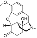

Fig. 12.2
Structure of oxycodone. “Oxycodone” by Fvasconcellos (talk contribs) – Own work (Licensed under Public Domain via Commons – https://commons.wikimedia.org/wiki/File:Oxycodone.svg#/media/File:Oxycodone.svg)
12.2.3 Hydromorphone
Hydromorphone is a semisynthetic opioid derived from morphine. Its structure is shown in Fig. 12.3. It functions primarily as a MOR agonist with less DOR activity. The time to maximum activity for oral administration is about 30 min and its duration of activity is about 4 h. Given intravenously, hydromorphone’s time to maximum activity is about 10–15 min, with the maximum effect at about 20 min and a duration of effect of about 4 h. It is metabolized in the liver to hydromorphone-3-glucurinode (H3G) (Trescot et al. 2008). H3G has no analgesic effect but can cause toxicity.
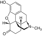

Fig. 12.3
Structure of hydromorphone. “Hydromorphone – Hydromorphon” by Yikrazuul – Own work (Licensed under Public Domain via Commons – https://commons.wikimedia.org/wiki/File:Hydromorphone_-_Hydromorphon.svg#/media/File:Hydromorphone_-_Hydromorphon.svg)
12.2.4 Fentanyl
Fentanyl is a synthetic opioid, as shown in Fig. 12.4. It is a MOR agonist. It undergoes rapid metabolism in the liver and cannot be given orally. It is available in IV, transdermal, transoral, mucosal, and inhaled formulations. Its time to onset after IV administration is less than a minute, with a peak onset time of about 5 min. However, it has a short half-life of 30–60 min. Transmucosal and inhaled fentanyl also have rapid onset of pain relief. Transdermal fentanyl has a 6–8 h of onset, and the pain relief equilibrates after several days. Subcutaneous fat acts as a reservoir for the transdermal fentanyl and can be active for 24 h after discontinuation of the transdermal drug. Fentanyl is metabolized in the liver to inactive and nontoxic metabolites (Trescot et al. 2008). With its rapid time of onset and short half-life without toxicity metabolites, it is the preferred drug for use with IV analgesia, including patient-controlled analgesia.


Fig. 12.4
Structure of fentanyl. “Fentanyl2DCSD” by Fuse809 (talk) – Own work (Licensed under Public Domain via Commons – https://commons.wikimedia.org/wiki/File:Fentanyl2DCSD.svg#/media/File:Fentanyl2DCSD.svg)
12.2.5 Methadone
Methadone is also a synthetic MOR agonist and is an antagonist of the N-methyl-D-aspartate (NDMA) receptor (see Fig. 12.5). This latter activity suggests it may be useful in neuropathic pain. It has a very complex and variable metabolism. It has a highly variable absorption, and so each patient must be titrated carefully. Also, although methadone has an analgesic duration of action of about 4–8 h, it has an elimination half-life of up to 150 h. Furthermore, methadone induces its metabolism enzymes in the liver, leading to increased methadone metabolism and drug requirements in the first week, but then it has stable metabolism. It is metabolized by the CYP3A4 enzyme, and thus it susceptible to multiple drug interactions, as described in the next section. Thus, it is hard to use at the end of life.
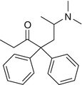

Fig. 12.5
Methadone. “Methadone” by Calvero – Selfmade with ChemDraw (Licensed under Public Domain via Commons – https://commons.wikimedia.org/wiki/File:Methadone.svg#/media/File:Methadone.svg)
12.2.6 Drug Interactions
Morphine and hydromorphone are metabolized through glucuronidation by the hepatic uridine diphosphate glucuronosyl transferase (UGT) enzymes with little to no P450 involvement. There are not many morphine and hydromorphone drug-drug interactions. Examples of possible interactions are with rifampin, ranitidine, and diclofenac (Armstrong and Cozza 2003).
In contrast, oxycodone and methadone are more prone to drug-drug interactions. Oxycodone is metabolized by the P450 CYP2D6 to oxymorphone, which is an FDA-approved commercially available analgesic. Inhibitors of P450 CYP enzymes have been implicated in oxycodone deaths. Strong inhibitors of CYP2D6 include selective serotonin reuptake inhibitors (SSRIs), such as fluoxetine, paroxetine, and ritonavir (Drummer et al. 1994; Jannetto et al. 2002; Jin et al. 2005) Dexamethasone also induces CYP2D6 and therefore could decrease the efficacy of oxycodone.
Methadone has a very high risk of drug-drug interactions because of its complicated metabolism to toxic and long-lived metabolites. P450 enzymes CYP3A4, CYP2B6, and CYUP2D6 are all involved in methadone metabolism. Curcumin, a popular herbal product, inhibits CYP2B6. In the same vein, grapefruit juice, noni, and pomegranate juice inhibit CYP3A4. Drugs inhibiting CYP3A4 include many antivirals; the azole antifungals including fluconazole, ketoconazole, and itraconazole; some macrolide antibiotics including erythromycin and clarithromycin; and some calcium channel blockers including verapamil and diltiazem. The CYP2D6 inhibitors were mentioned above.
12.2.7 Side Effects
The side effects of opioids are legion, but their efficacy usually outweighs their risks. The three general concerns with opioids in the dying patient are sedation, respiratory depression, and hypotension. Sedation can occur with starting and increasing doses of opioids, but it is usually temporary. Methylphenidate doses of 10–15 mg a day can reverse the sedation (Bruera et al. 1992; Wilwerding et al. 1995).
Respiratory depression can also occur, particularly with opioids that have toxic, long-lived metabolites such as methadone. The respiratory depression can be reversed with the use of naloxone. The half-life of naloxone after IV administration ranges from 30 to 80 min, which is shorter than most opioids. The patient must be observed for recurrence of the sedation. The recommended dose is from 0.4 to 2 mg, with further doses every 2–3 min if there is no response. This can be repeated after 20–60 min for recurrent sedation. Naloxone can also be given intramuscularly or subcutaneously. The lowest dose to reverse the sedation should be used, because the naloxone will also reverse the opioid’s analgesia, precipitating a pain crisis.
Cardiovascular effects, including hypotension, are uncommon, except with methadone. Hypotension can occur and appears to be related to histamine release by the opioids (Flacke et al. 1987). H1 blockade can decrease hypotension. This histamine release can also cause pruriitis, which is often mistaken for an allergic reaction. However, there is no mast cell release or IgA response to suggest an allergic component.
Another common, almost universal, side effect is constipation due to interaction with opiate receptors in the bowel inducing low motility. This is treated with fiber and other osmotic agents, stimulants, and hydration. If these are insufficient, then both methylnaltrexone and lubiprostone are effective for opioid-induced constipation (Ford et al. 2013). Both of these are very expensive. Misoprostol, an inexpensive prostaglandin E1 analog, is also effective, but a large percentage of patients cannot tolerate it due to cramping (Roarty et al. 1997; Soffer et al. 1994).
12.2.8 Conversion
The conversion from using one opioid to another is based upon general guidelines and are not based on any rigorous data. Further, there is incomplete cross-resistance between the opioids, and therefore the dose needs to be reduced anywhere from 25 to 50 % of the calculated conversion. Fentanyl cannot be converted from other opioids and must be titrated up from a low dose. Methadone is very difficult to convert to and from, and in the dying patient it may not be worthwhile to convert a patient to methadone, although it may be useful as an adjuvant if the patient is not responding to other opioids (Table 12.1 ).
Table 12.1
Properties of common opioids
Analgesic | Equivalent dosage | Time to maximum effect | Duration of analgesia |
|---|---|---|---|
Morphine, IV, IM | 10 mg | 5–10 min | 3–4 h |
Morphine, PO | 30 mg | 30–60 min | 3–4 h |
Oxycodone, PO | 20 mg | 10–15 min | 3–4 h |
Hydromorphone, IV, IM | 1.5 mg | 10–15 min | 3–4 h |
Hydromorphone, PO | 7.5 mg | 15–30 min | 2–3 h |
Fentanyl, IV | 0.05–0.1 mg | <1 min | 1–2 h |
Methadone, PO | Utilize special tables | ||
12.3 Nausea
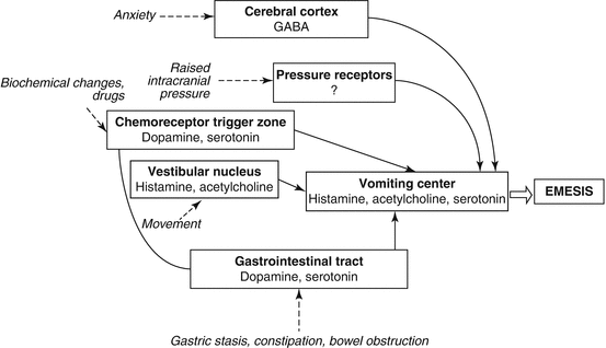
Nausea and/or vomiting occurs frequently in terminal cancer patients. It can arise from mechanical problems, such as constipation, esophageal obstruction or malignant bowel obstruction, metabolic toxicities such as renal failure or hepatic failure, from treatments such as with opioids or other medications, or from unidentified or unidentifiable causes (see Fig. 12.6). Obviously, correction of an identified cause such as a metabolic imbalance or medication problem should be the initial step to control nausea and vomiting.
12.3.1 Malignant Bowel Obstruction
A potentially correctable cause of nausea and vomiting in cancer patients is malignant bowel obstruction. High-volume vomiting with minimal odor and with brief episodes of cramping pain is often secondary to small bowel obstruction, whereas small-volume, foul-smelling emesis with more severe colic may represent colonic obstruction (Laval et al. 2014). Malignant bowel obstruction is more common with gynecological and gastrointestinal cancer. Obstruction can be from focal blockage of the bowel, such as from a primary colorectal cancer, or from peritoneal carcinomatosis. Radiographic evaluation can assist with diagnosis. For a patient that presents with abdominal pain, distention, nausea, and constipation, a plain abdominal film or CT scan may show complete or high-grade partial small bowel obstruction with proximal dilated loops of bowel with multiple air-fluid levels (Silva et al. 2009). Peritoneal carcinomatosis may present on CT with ascites, invasion of the fat of the greater omentum with streaking or nodules, invasion of the mesentery with nodules or anomalous fixation of the small intestine, and tumor implants on the walls of the intestine. Diffuse carcinomatosis may not be too small to be imaged (Diop et al. 2014).
Laval and colleagues from the French Society for Palliative Care published guidelines for the treatment of bowel obstruction from peritoneal carcinomatosis (Laval et al. 2014). They state that CT scanning is the gold standard for diagnosis, with a sensitivity of more than 90 %. Once clinical features and imaging confirm obstruction secondary to carcinomatosis, the patient can be evaluated for surgery. Surgery would rarely be recommended for a patient at the end of life. The role of stents in peritoneal carcinomatosis is also limited due to the usually diffuse nature of the obstructions and expected progression elsewhere. Placement of a venting gastrostomy tube is a low-morbidity procedure that can improve the quality of life of patients with obstruction. In a series reported by Issaka et al., there was successful placement of the venting tube in 95 of 96 patients (Issaka et al. 2014). The venting gastric tube provided complete relief from nausea and vomiting in 91 % of the patients.
Medical management of the symptoms of malignant obstruction often includes corticosteroids (Laval et al. 2014). Although the corticosteroids are generally well tolerated, their efficacy is also limited (Feuer and Broadley 1999). Anticholinergics, such as scopolamine and hyoscine butylbromide, act as antisecretory agents to reduce the volume of fluid in the bowels. These drugs have the common anticholinergic side effects such as dry mouth, tachycardia, and agitation. Glycopyrrolate, also an anticholinergic, has little penetration into the central nervous system, and so has fewer side effects (Soriano and Davis 2011). Octreotide can also be given, but at much greater cost. Opioids can be given for the abdominal pain, but opioids can also stimulate the bowel, causing more pain. In this case, they can be combined with the anticholinergics to reduce the colic (Soriano and Davis 2011).
12.3.2 Pharmaceutical Management
Davis, Walsh and colleagues at the Cleveland Clinic published their approach to the treatment of nausea (Gupta et al. 2013). They use a standard sequential approach to antiemetics. First, they use metoclopramide (a dopamine D2 antagonist) or haloperidol (a dopamine D2 antagonist), and then use olanzapine (an atypical antipsychotic medication with dopamine, serotonin, alpha-adrenergic, and cholinergic receptor activity) or chlorpromazine (a dopamine D2 antagonist) as a second-line treatment, and ondansetron (a selective 5HT3 receptor antagonist) as third-line treatment. For patients with ascites, a paracentesis can help relieve nausea. For patients with cerebral brain edema, they use dexamethasone. They recommend gabapentin (or carbamazepine) for relief of nausea from leptomeningeal carcinomatosis, but there are no clinical trials of its use (Gupta et al. 2013). Another approach to choosing an antiemetic treatment is mechanistic, that is, choosing the treatment based on the “emetic pathway” and cause of the nausea (Glare et al. 2011). However, there is no evidence that this approach is more successful than an empirical approach. The article by Glare and colleagues gives an extensive discussion of the medications that can be used for the treatment of nausea and vomiting.
Stay updated, free articles. Join our Telegram channel

Full access? Get Clinical Tree


