System
Variables
1 pt
2 pts
3 pts
RENAL
Radius (maximal diameter)
R ≤ 4 cm
4 < R ≤ 7 cm
R ≥ 7 cm
Low complexity: 4–6
Exo-/endophytic
≥50 %
<50 %
Endophytic
Moderate complexity: 7–9
Nearness to collecting system/sinus
N ≥ 7 mm
4 < N < 7 mm
≤4 mm
High complexity: 10–12
Anterior/posterior: a, anterior; p, posterior; x, not determined
–
–
–
h suffix if mass touches the renal artery/vein
Location relative to polar line
Entirely above or below polar line
Crosses polar line
>50 % of mass is across polar line or mass crosses the axial midline, or mass is between polar line
PADUA
Tumor size
≤4 cm
4.1–7 cm
>7 cm
Low complexity: 6–7
Renal sinus involvement
Not involved
Involved
–
Moderate complexity: 8–9
Collecting system involvement
Not involved
Involved
–
High complexity: ≥10
Exophytic rate
≥50 %
<50 %
Endophytic
Polar location
Superior/inferior
Middle
–
Tumor rim location
Lateral
Medial
–
DAP
Diameter of tumor
<2.4 cm
2.4–4.4 cm
>4.4 cm
Axial distance
>1.5 cm
≤1.5 cm
Overlap
Polar distance
>2 cm
≤2 cm
Overlap
Zonal NePhRO
Nearness to collecting system
Mass touches cortex
Mass touches medulla
Mass touches collecting system or crosses renal sinus
Low risk: 4–6
Physical location
Lower pole below collecting system
Lateral to but not touching collecting system
Upper pole or touches collecting system
Intermediate risk: 7–9
Radius of tumor (diameter)
<2.5 cm
2.5 ≤ R < 4 cm
≥4 cm
High risk: 10–12
Organization (exo-/endophytic)
>50 % exophytic
50 % endophytic 75 % exophytic
>75 % endophytic
C-index score:
Centrality index scoring
No point system but calculation of centrality index by the following:
0 = mass concentric to kidney center
Cross-sectional imaging and Pythagorean theorem to calculate distance from tumor center to kidney center. Division with tumor size to obtain centrality index
1 = periphery touching kidney center
Larger index = increased distance to kidney center
8.4.3 Radical Nephrectomy
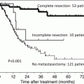
Complete excision by PN is preferred for SRMs in healthy individuals. In 2009, the American Urological Association (AUA) presented guidelines for clinical T1 renal masses listing radical nephrectomy (RN) as a viable treatment option for patients where PN is not technically feasible. In a multi-institutional study (EORTC 30904) [29], patients randomized to either nephron-sparing surgery (NSS) or RN for renal tumors ≤5 cm showed that the 10-year overall survival (OS) was 75.7 % for NSS compared to 81.1 % for RN (p = not significant). The 10-year progression rate for NSS was 4.1 % and for RN was 3.3 % (p = 0.48). The NSS group had a slightly higher rate of complications with pleural and splenic injury, bleeding, and urine leaks. Approximately 85.7 % of patients who underwent RN had renal dysfunction with an estimated glomerular filtration rate (eGFR) <60, compared to 64.7 % of patients after NSS at a median follow-up of 6.7 years. For advanced kidney disease (defined as eGFR <30), 10 % of RN and 6.3 % of NSS patients reached this point, and about 2 % of patients in each group demonstrated extreme renal dysfunction (eGFR <15). Thus, the decreased moderate renal dysfunction seen with NSS did not demonstrate a survival benefit in this group of patients for this follow-up time period.
The removal of the whole kidney for a peripherally located/exophytic SRM theoretically seems to remove an excess amount of normal kidney parenchyma unnecessarily. The surgeon should consider patient age and comorbidities, life expectancy, and oncologic goals of treatment when considering RN versus a nephron-sparing approach. Although, EORTC 30904 did not demonstrate a survival benefit in patients with clinical T1 masses who had NSS during follow-up, the higher rates of moderate renal dysfunction in RN patients may increase progressive renal insufficiency requiring dialysis along with its associated risk factors such as cardiovascular events after longer follow-up time periods [24, 25]. From a technical approach, RN for SRMs can be performed by laparoscopic or open surgery with similar steps as described above for PN with hilar vessel control and division. In general, RN should be reserved for masses whereby NSS is not easily possible and where the expeditious removal of the kidney facilitates recovery in patients with marginal surgical candidacy.
8.5 Locally Advanced Disease
Surgical excision of locally advanced RCC requires careful planning, patient optimization, and coordination of medical specialists and urologic surgeons. As in SRMs, the oncologic goals of locally advanced RCC are identical, to provide the greatest survival benefit with palliation of clinical symptoms, with the lowest morbidity possible. The definition of locally advanced RCC is typically defined as ≥T3 in the absence of distant metastasis [37]. For the surgical excision of RCC with concomitant thrombectomy in M0 (nonmetastatic) patients, the reported median survival range from 35 to 116 months with the 5-year CSS ranging between 40 and 65 % [38, 39]. For metastatic and T4 disease, the 5-year CSS is significantly lower ranging between 6.5 and 19 % [39, 40]. In comparison, the natural course or untreated RCC with VT is rather dismal with a recent Surveillance, Epidemiology, and End Results (SEER) database study reporting a median survival time of 5 months and a 1-year DSS as 29 % [41]. In this study, patients are of advanced stage and of poor performance status prohibiting primary surgical treatment. However, a subset analysis of nodal and metastasis-free (N0, M0) patients in this study demonstrate a significantly longer median survival of 14 months. Thus, the complete excision of RCC with concomitant VT removal may significantly increase survival [42].
The surgical excision of locally advanced RCC is more invasive compared to techniques developed for SRMs with surgical maneuvers performed to optimize exposure and removal. Due to the varying scope of locally advanced RCC, open, laparoscopic, and robotic techniques have been described with transabdominal and retroperitoneal approaches with different types of surgical incisions such as midline, subcostal/bilateral chevron, flank, and thoracoabdominal incisions. The midline incision allows exposure to the affected renal hilum as well as the contralateral renal vasculature and when extended to the thorax, the retrohepatic inferior vena cava, and the cardiac vasculature. Similar exposure can be obtained with bilateral chevron with an extended midline/thorax incision and with a flank incision extended to the thorax (a thoracoabdominal incision). Hepatic mobility may be facilitated by transection of the left triangular and coronary ligaments to provide exposure to the retrohepatic IVC.
The surgical steps for excision of RCC with associated venous thrombus (VT) include the isolation of the renal hilum with control of the renal artery first. The renal vein, IVC, and the contralateral renal vein are isolated and sequentially clamped cephalad and caudal to the VT. The VT can be visualized and monitored for extraction using transesophageal echocardiography. The VT is extracted with a cavotomy en bloc with the affected renal vein. The IVC can then be reconstructed primarily, or if the diameter is less than 50 % of the original diameter, a graft can be utilized. The VT may also directly invade the IVC; in this instance, the IVC may be removed en bloc with reconstruction using vascular grafts as needed. For VT above the diaphragm and into the cardiac vasculature, cardiopulmonary (CPB) and venovenous (VVB) bypass may be used to facilitate VT removal. In general, VVB is not used for VT involving the right atrium and, due to the shorter bypass circuit, may provide shorter operative times compared to CPB.
8.6 Lymphadenectomy
The EORTC 30881 was a randomized trial examining therapeutic benefit of RN with and without lymph node dissection (LND) [43]. A total of 772 patients were selected for randomization with 383 patients in the LND group and 389 in the non-LND group. The majority (~70 %) of these patients were of lower-stage tumors (≤T2). Pathological analysis of the LND dissections revealed an absence of LN metastasis in 332 patients out of 346 (96 %). Palpably enlarged LNs during surgery did not demonstrate LN metastasis as the majority (80 %) were negative and only 1 % with non-palpable nodes were positive. In the patients that did not undergo a LND, 9 % of patients had enlarged LNs. These LNs were excised for staging purposes or biopsied with 12 % demonstrating LN metastasis. In all, 96 % of the resected group did not show LN metastasis, and there were no significant differences in all survival parameters (overall, time to progression, or progression-free survival) at a median follow-up period of 12.6 years. The main criticism of this study was that the majority of patients were of low-risk disease, and benefits of a formal LND would not demonstrate much of a survival benefit.
LND may be of limited benefit for low-stage renal tumors as noted previously. On the contrary, it is hypothesized that LND may benefit higher-stage tumors and/or renal tumors with adverse pathological features. As retrospective studies have shown LN metastasis to be stage dependent ranging between 12 and 37 % for T3–4 tumors [44, 45]. At our institution, the borders of a formal LND are ipsilateral hilar LNs and para-aortic LNs from the crus of the diaphragm to the aortic bifurcation for left-sided tumors. For right-sided tumors, the interaortocaval and para-caval LNs are removed from the crus of the diaphragm to the large vessel bifurcation [46].
8.7 Cytoreductive Nephrectomy
Approximately 25 % of RCC patients will initially present with metastatic disease with treatments mainly focused on systemic therapies [47]. In 2001, two phase III randomized clinical trials reported a statistically significant survival benefit when radical nephrectomy was combined with interferon-alpha2b. In EORTC 30947 [16], 42 participants were randomly assigned to the RN before interferon-alpha2b and 43 to the interferon-alpha2b alone. The time to progression was 5 months versus 3 months (HR 0.60, 95 % CI 0.36–0.97) and median survival of 17 versus 7 months (HR 0.54, 95 % CI 0.31–0.94) with favorable survival observed when combined with RN. The Southwest Oncology Group (SWOG) randomized 121 to interferon alone versus 120 to RN plus interferon-alpha2b [15]. When combined with surgery, there was a 3-month (P = 0.05) improvement in median survival (11.1 vs. 8.1 months), independent of performance status and site of metastatic spread.
Although these two trials used immunotherapies, they have continued to motivate cytoreductive nephrectomy (CN) in the contemporary targeted therapy era. Targeted agents such as the tyrosine kinase inhibitors (sorafenib, sunitinib, and axitinib), mTOR inhibitors, and VEGF antibodies have been explored as agents used after CN [48–53]. These studies examined CN in subgroup analysis with the primary end point of progression-free survival to show promising trends in survival improvement. There are two ongoing randomized trials accruing to examine CN with targeted therapies. The EORTC 30073 (SURTIME trial, NCT 01099423) is a randomized phase III trial comparing immediate versus delayed (after receiving two cycles of sunitinib) nephrectomy in patients with synchronous metastatic RCC. The trial is still accruing with an expected enrollment of 458 patients. The CARMENA trial (NCT 00930033) randomizes to RN and sunitinib versus sunitinib alone with the primary end point of overall survival. The estimated accrual is 576 patients. It is expected that the results of these two trials will refine the role and timing of CN with targeted agents.
8.8 Conclusion
The spectrum of renal masses from SRMs to locally advanced and metastatic disease varies the management from active surveillance to invasive procedures including surgery. Due to the variability in biology and relative resistance to systemic therapies of RCC, surgery remains an important component of treatment. Since the first modern description of radical nephrectomy for tumor was described in the late 1800s, refinements in surgical technique have evolved to remove the kidney, perinephric fat, and regional lymph nodes for primary oncologic control. With partial nephrectomy, the removal of the whole kidney is not necessary for SRMs, and the development of laparoscopy and robotic techniques have advanced the treatment paradigm. As patients present in different stages of disease each with their own unique clinical factors, informed counseling is paramount to meet their expectations. Furthermore, as many treatment methodologies are based on retrospective and observational studies, enrollment in clinical trials should be encouraged. As we await the conclusion of current trials with the introduction of new systemic therapies, the role of surgical excision is evolving.
Related posts:

Stay updated, free articles. Join our Telegram channel

Full access? Get Clinical Tree


