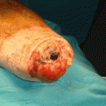
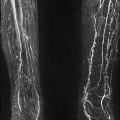
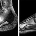
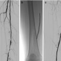
Figure 9.1
Pathway for revascularisation of the diabetic foot
Choice of Bypass Conduit
There are three choices when considering a conduit for bypass surgery. By far the superior choice is the patient’s own vein. Second are synthetic man-made grafts composed of either ring-supported “Dacron” or ePTFE. Both will usually be reinforced on the outside with spiral plastic supports to prevent kinking. A recent development are ePTFE grafts “rifled” on the inside to produce spiral flow of blood within the conduit, to reduce neointimal hyperplasia at the anastomosis and increase longevity. Although early results for these grafts are encouraging, no long-term data exists at present. The final option is cadaveric vein, which has been cryopreserved after harvest from a post-mortem donor. Cadaveric vein use in the UK has been limited, largely due to cost and the limited outcome data available.
A pre-operative duplex scan for vein mapping is essential as it allows assessment of the venous conduit quality (>3 mm is considered acceptable) and aids accurate intra-operative conduit harvesting, avoiding complications with skin flap necrosis. Vein is the preferred gold-standard conduit for bypass procedures as they have more durable patency rates and are less likely to suffer infection compared to prosthetic Dacron or ePTFE conduits. As well as the great saphenous vein and short saphenous vein, the basilic and cephalic arm veins can be harvested to good use.
The Inflow Vessel
The proximal inflow vessel must be as disease-free as possible and is usually the infra-inguinal common femoral artery, but it can be derived from the supra-inguinal external iliac or the infra-inguinal profunda femoris or superficial femoral artery. In diabetic patients it is often possible to perform shorter bypasses, using the popliteal artery behind the knee as an inflow vessel. This removes the need for a longer venous conduit required to perform femoral distal bypass, with equally good long-term results achieved for the shorter bypasses. In some patients, angioplasty and stenting of the iliac arterial segment may be required beforehand, to allow the use of the common femoral artery as the inflow vessel.
The Outflow Target Vessel
The distal outflow target vessel for graft anastomosis is typically the most disease-free tibial artery identified on angiography. Ideally it should cross the ankle into the plantar pedal arch to provide a realistic chance of ulcer healing. The distal outflow target vessel can be the popliteal artery above or below the knee or the best-quality infra-geniculate tibial artery crossing the ankle joint, which may or may not be in continuity with the plantar pedal arch in the foot. The nomenclature of lower limb bypass surgery reflects this.
Popliteal target – femoro-popliteal bypass
Tibial vessel target – femoro-distal bypass
Plantar pedal arch target – femoro-ultra distal bypass
Technical Considerations During Bypass Surgery
The small size of distal target arteries makes the anastomosis in femoral-distal bypass more technically challenging, with a greater chance of early failure. Wherever possible the most proximal landing zone in the target vessel should be used. Magnifying surgical eyewear e.g. Loupes, should be worn by the operating surgeon performing the distal anastomosis. This enables accurate, small, evenly-spaced suture bites to be taken and the identification of debris or small intimal flaps.
Prior to venous conduit harvesting, it is helpful to mark the course of the vein pre-operatively using ultrasound, to facilitate accurate skin incisions during vein harvest and to confirm the vein size (>3 mm in diameter ideally) and the quality of the vein (i.e. that it is free from thrombophlebitis). The great saphenous vein (GSV) is most commonly used and arises in the foot and passes anterior to the medial malleolus at the ankle, ascending the leg medially and superficial to the muscles within its own facial envelope, before diving deep in the groin to join the common femoral vein at the sapheno-femoral junction. The GSV is ‘harvested’ or disconnected from the venous system and used to carry higher pressure oxygenated arterial blood distally. Over time, the thin-walled GSV becomes ‘arterialised’ to the point that at revision surgery it can be difficult to tell a vein graft from a native artery.
Vein grafts are more infection-resistant and durable compared to prosthetic grafts. An infected graft often results in limb loss as revision surgery is often difficult and risky. Vein grafts do not develop the impervious bio-layer of bacteria that an artificial graft does, making antibiotic treatment feasible in the first instance. Occasionally the contralateral great saphenous vein or the basilic and cephalic veins in the arm are harvested as conduits in preference to prosthetic grafts. If an individual segment of vein is not of sufficient length to complete the bypass, two or three segments of vein can be harvested and ‘spliced’ together, to produce one long conduit.
Veins taper up in size from 2 to 3 mm at the ankle to around 8–10 mm at the sapheno-femoral junction in the groin, as more tributaries drain into them. They contain one-way valves that prevent blood returning to the foot under the effects of gravity, when a patient is upright and stationary. These two points must be borne in mind when deciding on how to anastomose the vein graft onto the arteries. If the vein is reversed in direction to counter the effects of the valves, a size mismatch occurs with a large diameter artery proximally but a small-diameter vein and vice-versa at the distal end. This can usually be corrected for in the popliteal segment, but more distally it can be technically challenging to join a 10-mm diameter vein graft to a 2-mm tibial artery. In these cases it may be preferable to leave the vein in-situ, passing a valvulotome instrument down the vein that cuts and destroys the valve leaflets, allowing reverse flow of blood within the vein. There are no differences in long term outcomes between reversed or in-situ vein techniques for bypass. An in-situ bypass may help to avoid a size mismatch between smaller target vessels and the venous conduit but may take slightly longer to harvest and prepare with a valvulotome. There is also a small risk of injury to the venous conduit as the valvulotome is passed. The authors recommend using an expandable valvulotome, which can prepare vessels as small as 1.5 mm in diameter.
Stay updated, free articles. Join our Telegram channel

Full access? Get Clinical Tree


