Fig. 16.1
Abdominal ultrasound images demonstrating a grossly dilated CBD (3 cm between yellow cross-hairs) (upper left and lower left images). Duplex colour-flow imaging of the portal vein (blue) and hepatic artery (red) adjacent to the dilated CBD (upper left image). Centrally dilated intrahepatic ducts were also seen tapering within the hepatic parenchyma (upper right and lower right images). CBD common bile duct; LLL left lobe of liver
These ultrasound findings raised the possibility of a choledochal cyst, and therefore the patient proceeded to magnetic resonance cholangiopancreatography (MRCP). This demonstrated centrally dilated intrahepatic ducts which tapered to normal calibre beyond the secondary order division. The cystic duct, CHD, and CBD were all dilated, with gross fusiform dilatation of the proximal CBD up to a maximum diameter of 3.5 cm (Fig. 16.2, and supplemental video).
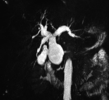
Fig. 16.2
Representative image of the 3D cholangiography reconstruction from the MRCP. The full reconstruction can be viewed online (A supplementary video has been submitted)
The pancreaticobiliary segment of the common duct was long, with the pancreaticobiliary junction identified proximal to the usual site. There was no sign of intrahepatic or extrahepatic ductal calculi, no evidence of intrahepatic or extrahepatic strictures, and no other abnormalities in the liver parenchyma, spleen, kidneys, adrenals, or pancreas. These findings confirmed the diagnosis of a Type IVa choledochal cyst.
Diagnosis and Assessment
Choledochal cysts are rare developmental malformations of the biliary tree, comprising 1% of all benign biliary conditions [1]. The classical presentation is of a female child with abdominal pain, jaundice, and an abdominal mass, although up to a quarter of patients present as adults [2]. Perhaps due to the increasing availability of high-quality noninvasive imaging, patients are more frequently being diagnosed in adulthood [3].
An abdominal ultrasound would be the commonest initial investigation for patients with abdominal pain and deranged liver function tests, and this test is sensitive for the detection of intrahepatic cystic disease. Delineating the extent of choledochal cystic disease is best accomplished by magnetic retrograde cholangiopancreatography MRCP [4]. A triple-phase contrast-enhanced CT can be complementary in determining the relationship of extrahepatic cystic disease to neighbouring vascular structures. If MRI is contraindicated, direct cholangiography either by endoscopic retrograde cholangiography (ERC) or percutaneous transhepatic cholangiography (PTC) is recommended, but this is associated with a greater risk to the patient.
Our own experience is that patients commonly present with abdominal pain, and most have at least one episode of cholangitis, with many having intermittently deranged liver function tests.
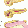
The modified Todani classification is the most widely established in clinical practice, and segregates patients on the anatomical distribution of the abnormalities (Fig. 16.3) [5]. Across published series, approximately 80% of patients have type I cysts, 15% have type IVa cysts, and the remainder are classified as types II, III, IVb, and V [6]. Type IVa cysts are more common in adults.
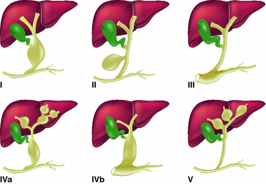
Fig. 16.3
Todani classification of choledochal cysts, adapted from [5]. Type I solitary extrahepatic cyst (can be subclassified according to fusiform (If) or cystic (Ic) type); Type II extrahepatic bile duct diverticulum; Type III cyst of the common pancreaticobiliary channel (choledochocele); Type IVa extrahepatic and intrahepatic cysts; Type IVb multiple extrahepatic cysts; Type V intrahepatic cysts (Caroli’s disease)
Clinical Pearls
Cyst drainage procedures (e.g. cyst-duodenostomy) have poor long-term success rates due to complications with the cyst remnant and should be avoided in favour of primary extrahepatic cyst excision and biliary reconstruction.
The risk of cholangiocarcinoma remains for all patients with choledochal cysts even after cyst excision and this appears higher in patients with an APBJ or residual intrahepatic disease. These patients should remain under long-term follow-up. The frequency and mode of follow-up are largely down to surgical preference but those patients developing cholangiocarcinoma following cyst excision currently have a poor prognosis.
Incidence and Aetiology
Choledochal cysts are rare globally, with an estimated incidence of 1 in 100,000 live births in Western countries, but they are more common in Asian countries and up to 100-fold more frequent in Japan [7]. Several studies report a strong association between an abnormal pancreatico-biliary junction (APBJ) and choledochal cysts [8, 9]. A report of monozygotic twins provides further support of the direct association. One sibling presented at 2 years of age with abdominal pain and abnormal liver function tests, and was demonstrated to have an anomalous pancreatico-biliary junction and associated type I choledochal cyst. The other twin was asymptomatic and had a normal pancreaticobiliary junction and biliary tree [10].
Reflux of pancreatic exocrine secretions into the biliary tree due to an APBJ has been proposed as the central aetiological factor in choledochal cyst development. The finding of a long common pancreaticobiliary duct in our case further supports the association of APBJ and choledochal cysts and the long common channel hypothesis [11].
The in utero finding of a choledochal cyst prior to the development of the exocrine pancreas, however, casts doubt on the necessity of reflux of exocrine secretions in cystic degeneration, and an alternative hypothesis is that neonatal biliary obstruction is the central factor [12].
Clinical Course
Regardless of the underlying aetiology, choledochal cysts are associated with multiple complications. Impaired biliary drainage and cholestasis predispose to stone development, and infectious sequelae are common, including cholecystitis, cholangitis, and hepatic abscess [13]. Pancreatitis is also common and may be secondary to stones, causing pancreatic duct obstruction, a direct result of the APBJ, or as a consequence of ERCP undertaken for biliary drainage [14]. Recurrent cholangitis and biliary obstruction can lead to cirrhosis and portal hypertension, and patients should be thoroughly assessed for these complications prior to operative intervention [13, 15, 16].
Up to 75% of patients with type I cysts have ductal stones, and the prevalence is even higher in patients with type IVa cysts [17]. It is important to establish the extent of intrahepatic disease and the presence of abscesses, stones, or strictures pre-operatively, as these pathologies may not be adequately treated by hepaticojejunostomy and may result in recurrent stone formation and sepsis [17].
Patients with choledochal cysts have up to a 100-fold higher risk of developing cholangiocarcinoma than the general population [18]. The incidence of malignancy increases with age, with the majority of cancers presenting by the fourth decade of life. The overall incidence of malignancy is 11%, [2, 19, 20] and those patients with type I or type IVa cysts appear to be at highest risk [21, 22].
The risk of cholangiocarcinoma remains following resection of extrahepatic cystic disease, with a 10% reported cumulative incidence in one Japanese series up to 25 years post-excision, although the risk appears lower in Western populations [18, 23]. Patients at higher risk include those with an APBJ or those with incomplete cyst excision, such as patients with type IVa cysts and residual intrahepatic disease [22].
A key principle in the treatment of choledochal cysts is complete excision of the extrahepatic disease, as patients who have undergone a cyst drainage procedure—such as transduodenal sphincteroplasty, choledochoduodenostomy, or choledochojejunostomy—have a higher risk of cholangiocarcinoma than those patients who have undergone cyst excision [23, 24].
Interestingly, drainage procedures in patients without choledochal cysts are also associated with an increased risk of cholangiocarcinoma, and this may reflect the carcinogenic effect of chronic enteric reflux on the biliary epithelium [25].
Alternative Approaches and Controversies
Patients with Type IVa choledochal cysts have both intra and extrahepatic disease. One approach is to only resect the extrahepatic disease. An alternative is to resect both the intrahepatic and extrahepatic components, with a greater risk of post-operative morbidity but potentially a reduced risk of future intrahepatic complications.
Our approach is to undertake excision of the extrahaptic disease only, reserving synchronous resection for selected patients with predominantly unilobar disease and evidence of intrahepatic strictures or multiple intrahepatic stones at high risk of recurrent biliary sepsis.
Operative Management
Our patient underwent laparotomy with intraoperative ultrasonography, confirming dilated intrahepatic and extrahepatic ducts with no evidence of ductal stones, liver abscess, or other parenchymal lesions. The extrahepatic biliary tree was excised from the confluence of the hepatic ducts to the superior border of the pancreas with an en-bloc cholecystectomy. On-table choledochoscopy was performed and confirmed no intrahepatic ductal stones or strictures (See Technical Elements box below).
A Roux-en-Y hepaticojejunostomy was performed for reconstitution of biliary drainage. This would be our favoured approach for both type I and type IVa choledochal cysts, and is increasingly recognised as the established standard in the literature [26]. Cyst excision and reconstruction should be performed in all cases, including those patients who have previously undergone a primary cyst drainage procedure, as post-operative complication rates are low and longer term outcomes are superior [24]. If the gallbladder is present, a cholecystectomy should always be performed as part of the procedure [27].
Our practice would be to perform the biliary-enteric anastomosis at the confluence of right and left hepatic ducts. Spatulation of the left hepatic duct allows a wide anastomosis to be constructed in most patients, reducing the risk of a post-operative anastomotic stricture and therefore reducing the need for revisional surgery [28]. If the intrahepatic component of the choledochal cyst involves the confluence of the hepatic ducts, we would still recommend excision of the extrahepatic component and anastomosis to an epithelial-lined portion of the intrahepatic cyst. In patients with distal extrahepatic disease, complete excision of the extrahepatic ducts and anastomosis at the biliary confluence should still be performed.
Related posts:
 Resection of Centrally Located Cystadenoma/Cystadenocarcinoma
 Debulking of Extensive Neuroendocrine Liver Metastases
 Pancreatic Adenocarcinoma in the Neck of the Pancreas Involving the Celiac Trunk (Appleby Procedure)
 Gallbladder Cancer with Common Bile Duct Invasion
 Chronic Pancreatitis: Puestow and Frey Procedures
 Resection of Renal Cell Carcinoma Involving the Liver with Tumor Thrombus Extending into Inferior Vena Cava Requiring Venovenous Bypass
Stay updated, free articles. Join our Telegram channel

Full access? Get Clinical Tree


