The outcome of patients with stage I nonseminomatous germ cell tumors (NSGCTs) is excellent, with 99% overall 5-year survival. If treated with orchiectomy alone, approximately 30% of patients relapse with metastatic disease. Therefore adjuvant therapy with either chemotherapy or retroperitoneal lymph node dissection (RPLND) is often recommended, despite impressive results with active surveillance alone. This article addresses the risks and benefits of each approach.
An increasing proportion of patients with germ cell tumors are presenting with stage I disease. This trend may be partly because of increased awareness among young men. Stage I seminoma occurs more frequently than nonseminoma; however, the unifying factor for both conditions is the excellent outcome after orchiectomy irrespective of subsequent treatments. For example, in a recent study with 740 patients with stage I nonseminomatous germ cell tumors (NSGCTs) showed no NSGCT-related deaths. This finding underlines the challenging nature of prospective randomized studies in this rare disease, because current standard treatment results in disease-specific survival of nearly 100%.
The treatment options after orchiectomy include adjuvant chemotherapy, retroperitoneal lymph node dissection (RPLND), and surveillance. These options have all become accepted standards of care, based predominantly on numerous single-arm descriptive studies ( Tables 1–3 ). The lack of randomized data favoring one treatment has resulted in standard treatment pathways differing among institutions and countries, with Europe leaning toward surveillance and adjuvant chemotherapy and North America recommending surveillance or RPLND.
| Study (y) | Number of Patients | Stage II at Pathology | Relapse Rate for Pathology Stage I a | Relapse Rate for Pathology Stage II a | Overall Survival |
|---|---|---|---|---|---|
| Donohue et al, 1993 | 379 | 112 (30%) | 12% | 34% | 99% |
| Hermans et al, 2000 | 292 | 66 (32%) | 10% | 23% | NA |
| Albers et al, 2003 | 182 | 62 (28%) | 18% | 25% | NA |
| Nicolai et al, 2004 | 322 | 60 (19%) | NA | NA | 99% |
| Stephenson et al, 2005 | 309 | 91 (30%) | 7% | 34% | 99% |
a This includes only those patients who did not go on to receive post-RPLND adjuvant chemotherapy.
| Study (y) | Number of Patients | Years of Follow-Up | Relapse Rate | Overall Survival a |
|---|---|---|---|---|
| Read et al, 1992 | 373 | 5 | 100 (27%) | 98% |
| Colls et al, 1999 | 248 | 4.5 | 70 (28%) | 98% |
| Francis et al, 2000 | 183 | 5 | 52 (28%) | 99% |
| Daugaard et al, 2003 | 349 | 5 | 86 (29%) | 100% |
| Ernst et al, 2005 | 194 | 4.5 | 57 (29%) | 100% |
| Oliver et al, 2004 | 234 | 3.0 | 71 (30%) | 98% |
| Sturgeon et al, 2011 | 371 | 6.3 | 104 (28%) | 99% |
| Study (y) | Number | Chemotherapy Regimen | Number of Cycles | Years of Follow-Up | Relapses | Testis Cancer Deaths |
|---|---|---|---|---|---|---|
| Cullen et al, 1996 | 116 | BEP | 2 | 4 | 2 (2%) | 2% |
| Oliver et al, 2004 | 28 | BEP | 2 | 3 | 1 (4%) | 4% |
| Chevreau et al, 2004 | 40 | BEP | 2 | 10 | 0 (0%) | 0% |
| Pont et al, 1996 | 42 | BEP | 2 | 6 | 2 (5%) | 3% |
| Tanstad et al, 2009 | 157 | BEP | 1 | 5 | 5 (3%) | 0% |
| Oliver et al, 2004 | 47 | BEP | 1 | 3 | 3 (7%) | 0% |
| Klepp et al, 2003 | 57 | PVB | 2 | 3 | 1 (2%) | 0% |
| Dearnley et al, 2005 | 115 | BOP | 2 | 6 | 2 (2%) | 1% |
Most patients with stage I NSGCTs will not experience relapse after orchiectomy. Therefore, adjuvant treatment in most individuals is without benefit and potentially harmful, making a stratified approach to treatment based on the identification of risk factors associated with relapse an attractive prospect. The two factors that are most consistently associated with relapse include lymphovascular invasion and of embryonal carcinoma at histology. However, even with this knowledge, a group with a greater than 50% risk of relapse is difficult to identify.
Finally, this article addresses survivorship issues associated with stage I NSGCT. Given that cure rates approach 100%, decisions about treatment must take short- and long-term toxicity into account. Surgery and chemotherapy both have established short-and long-term risks, which range from bowel obstruction and loss of antegrade ejaculation after RPLND to the development of cardiovascular disease and second cancers after chemotherapy. These factors militate in favor of surveillance. However, even the surveillance group is exposed to risk, particularly from the radiation exposure associated with repeated cross-sectional imaging, which can occur up to 12 times over a 5-year period.
Clinical stage I disease
Clinical stage I disease is defined as no evidence of disease on CT of the chest, abdomen, and pelvis and normal serum tumor markers (α-fetoprotein [AFP], human chorionic gonadotropin [HCG], and lactate dehydrogenase [LDH]). Patients with persistently elevated makers after orchiectomy are considered to have metastatic disease, and chemotherapy is recommended. The results for RPLND in patients with elevated serum tumor markers have been disappointing because relapses outside of the surgical field have been frequent.
The retroperitoneal lymph nodes are the commonest site (>70%) of metastatic spread in NSGCTs. Lymph nodes larger than 1 cm are considered abnormal. However, borderline lymph nodes 8 to 10 mm in the primary landing zones may be worthy of more frequent investigation or intervention. These primary landing zones include the paracaval, precaval, interaortocaval and preaortic nodes for right-sided tumors, and the para-aortic, preaortic, and interaortocaval nodes for left-sided tumors.
Understaging of the retroperitoneum with CT occurs in 25% to 30% of patients, even with modern scanners. 18-Fluoroudeoxyglucose–positron emission tomography (FDG-PET)/CT has not helped further characterize these nodes and cannot be recommended in the routine staging for this disease. RPLND gives more accurate data than CT scanning regarding retroperitoneal involvement. However, RPLND is associated with morbidity and is not considered necessary to complete staging.
Prognostic factors for stage I NSGCT
During the 1980s, an investigation of more than 250 patients with stage I NSGCT showed several histologic factors that predisposed to relapse, including tumor invasion of testicular veins, lymphatic invasion by the tumor, absence of yolk sac elements, and the presence of embryonal cell carcinoma within the cancer. The absence of any of these factors resulted in a 100% 3-year relapse-free survival, whereas the presence of three or four factors was associated with a 58% relapse rate over the same period. These data were successfully externally validated in a larger cohort.
Subsequent investigators simplified the prognostic index through combining the presence of venous and lymphatic invasion as only one prognostic factor. This factor, along with the proportion of the tumor that consisted of embryonal carcinoma, became the most important factors over the next 2 decades. However, positive predictive values for relapse in high-risk groups remain less than 70%.
A more recent retrospective analysis of 23 publications between 1979 and 2001 investigated prognostic factors in more than 2500 patients with stage I NSCGT. Lymphovascular invasion remained the most significant prognostic factor (odds ratio [OR], 5.2; 95% CI, 4.0–6.8). Other factors, such as the presence of embryonal carcinoma in the primary tumor (OR, 2.9; 95% CI, 2.0–4.4) and a high pathologic stage of the tumor (OR, 2.6; 95% CI, 1.8–3.8), were also of significance.
Other makers such as MIB-1, p53, bcl-2, cathepsin D, and E-cadherin have been investigated in this setting with mixed results, although they have not been established in routine use.
RPLND for stage I NSGCT
Except for choriocarcinoma, NSGCTs follow a predictable pattern of metastasis. The landing zones within the retroperitoneum are the most commonly affected sites. RPLND not only accurately differentiates between stage I and II disease but also significantly reduces the probability of systemic relapse. Relapse within the retroperitoneal field after RPLND occurs in fewer than 1% of patients at centers of excellence, and overall survival is roughly 99% at 5 years. This approach appears safe and successful, with relatively few long-term side effects (retrograde ejaculation being the notable exception). RPLND also has the potential advantage of removal of chemotherapy-resistant teratoma, which is present in 30% of patients with stage II disease. Surgical removal of teratoma is required because it can undergo malignant transformation or become inoperable. The absence of teratoma in the primary tumor does not equate with absence in the retroperitoneal lymph nodes, making who is most likely to benefit from surgery difficult to predict. A major downside to RPLND is that relapse can occur outside the retroperitoneum in approximately 5% to 10% of patients with pathologic stage I disease.
RPLND for clinical stage I disease is safe with relatively few long-term side effects. Series published from Indiana University and Cleveland Clinic reported no perioperative deaths or permanent disability. Major complications were reported in only 2% to 3% of patients. The most significant long-term side effect with RPLND is the loss of antegrade ejaculation, which occurs in fewer than 5% of patients whose surgery is performed by a highly experienced surgeon. Other complications, which occur in approximately 1% of patients, include small bowel obstruction, wound infections, or lymphocele. It has been speculated that these excellent results are partly from the experience of the centers and the high volume of surgery, because most series have been reported by high-volume centers. These results seem to not be reproducible in the community setting.
Randomized prospective data from the German group compares one cycle of bleomycin, etoposide, platinum (BEP) with RPLND for treating clinical stage I disease. Both cancer centers and community-based hospitals participated, and 61 centers accrued a total of 382 subjects. The results showed that after a median follow-up of more than 4 years, none of the patients had died of germ cell tumors. However, 15 patients had experienced recurrence (2 in the chemotherapy arm vs 13 in the RPLND arm; P <.005). The 13 recurrences in the RPLND arm were observed within 17 months of therapy, with 5 (3%) of these relapses occurring in the RPLND field.
A modified ipsilateral nerve-sparing RPLND was allowed in this study, which may account for the relatively high relapse rate within the retroperitoneum (3%). It is also of note that such a low relapse rate was achieved with a single cycle of BEP chemotherapy, which is not generally considered chemotherapy standard of care. Overall, this study confirmed that chemotherapy results in a lower relapse rate than RPLND and supported the position that RPLND is best performed in specialist centers rather than in community hospitals.
Surveillance for stage I NSGCT
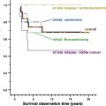
Surveillance after orchiectomy is an attractive option, particularly in the absence of high-risk features, when the chances of relapse are less than 20% (see Table 2 ). It is also an option in the high-risk setting because only approximately 50% of these patients relapse. Overall survival for surveillance is indistinguishable from the results seen with adjuvant treatments (98%–100%), resulting in some institutions pursuing a surveillance program for all patients with stage I NSGCT. When relapses occur, they do so in predicable anatomic locations and mostly within 2 years of orchiectomy (median time to relapse, 7 months). Late relapses in patients with stage I NSGCT undergoing surveillance are uncommon, occurring in 0% to 3% of men. Serum tumor markers are frequently elevated in patients with metastatic NSGCT, facilitating the detection of relapse and reducing the dependence on imaging studies. Together these factors make surveillance an attractive option for potentially all patients.
Cross-sectional imaging with CT identifies 48% to 53% of relapses, whereas raised serum tumor makers are responsible for indentifying 29% to 39% of relapses in these patients. Therefore, guidelines regarding the follow-up of these patients tend to focus on frequent cross-sectional imaging over the first 2 years. Subsequent imaging between years 2 and 5 varies depending on the treatment center. Serum tumor makers are measured regularly during this period. Guidance for follow-up varies a great deal depending on the individual institution and required clarification. Some recommend cross-sectional imaging every 3 to 6 months for up to 5 years, whereas others reduce the frequency of imaging and stop this follow-up much sooner. A prospective study compared two and five CT scans over the first 2 years of follow-up after orchiectomy. The results showed that a less-frequent imaging protocol did not predispose to an increased risk of relapse with more advanced disease. In view of the risk associated with radiation exposure, it seems wise to follow the guidance of this study to perform fewer scans.
The main concern with surveillance is the comparatively high risk of relapse, especially in high-risk patients. Waiting until relapse also results in a significant proportion of patients requiring RPLND after chemotherapy, because many patients do not experience a radiologic complete remission with chemotherapy for metastatic disease. Teratoma (40%) and viable tumor (10%) are frequently seen at surgery in this setting.
Overall surveillance seems safe and very attractive, provided that patients comply with follow-up and cross-sectional imaging protocols. The lack of randomized data in this area should not discourage clinicians from pursuing this approach, especially in patients with low-risk disease. Recent data suggest that a less-frequent cross-sectional imaging may not have a detrimental effect on relapse.
Stay updated, free articles. Join our Telegram channel

Full access? Get Clinical Tree


