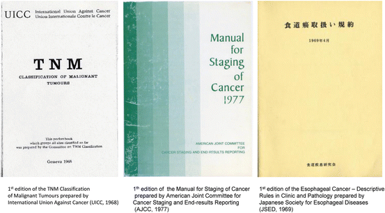
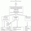
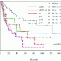
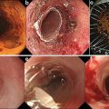
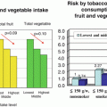
Fig. 5.1
History of TNM Classification of Malignant Tumours, AJCC Cancer Staging Manual and Japanese Classification of Esophageal Cancer, TNS* Tumor Nomenclature and Statistics, CSCAS* Clinical Stage Classification and Applied Statistics
In 1933, the International Union Against Cancer—Union Internationale Contre le Cancer (UICC)—was established as a nonprofit and non-government organization.
The TNM system for the classification of malignant tumors was developed by Pierre Denoix (France) in the 1940s. In 1950, the UICC appointed a Committee on Tumor Nomenclature and Statistics and agreed general definitions for the local extension of malignant tumors. In 1954, the Research Committee of the UICC set up a special Committee on Clinical Stage Classification and Applied Statistics to extend the general technique of classification to cancer at all sites.
Between 1960 and 1967, the Committee published nine pamphlets describing proposals for the classification of 23 sites. In 1968, these pamphlets were combined into a booklet, which was substantially the 1st edition of the TNM Classification of Malignant Tumours (Fig. 5.2) [1].
In 1995, the project started to publish Prognostic Factors in Cancer, a compilation and discussion of the prognostic factors in cancer, both anatomic and nonanatomic, at each of the body sites. The latest 7th edition of TNM Classification [7] contains rules of classification and staging that correspond with prognostic factors appearing in the 7th edition of the AJCC Cancer Staging Manual (2009) [14].
In 2010, the name of the UICC was changed to the Union for International Cancer Control.
5.2.2 A History of the AJC/AJCC and the AJCC Cancer Staging Manual (Fig. 5.1) [1–26]
The American Joint Committee for Cancer Staging and End Results Reporting (AJC) was first organized in 1959. The founding organizations of the AJC were several scientific societies including the American College of Surgeons and the National Cancer Institute. In 1976, the AJC sponsored a National Cancer Conference on classification and staging. The deliberation at this conference led to the development of the 1st edition of the Manual for Staging of Cancer which was published in 1977 (Fig. 5.2) [8].
In 1980, the new name, the American Joint Committee on Cancer (AJCC), was selected. Since the early 1980s, the close collaboration of the AJCC and the UICC has resulted in uniform and identical definitions and stage groupings of cancers for all anatomical sites. Initially in the 3rd edition of the Manual for Staging of Cancer (AJCC, 1988) [10] and the 4th edition of the TNM Classification of Malignant Tumours (UICC, 1987) [4], and since then the same TNM categories and stage grouping for esophageal cancers have been presented by both organizations.
Since the 1990s, the TNM staging of cancer has become widely adopted throughout the USA, and the terminology in the AJCC-TNM system is used for cancer reporting. Since the 5th edition published in 1997 [12], the new name, the AJCC Cancer Staging Manual, has been used.
5.2.3 A History of the JSED/JES and the Japanese Classification (Fig. 5.1) [1–27]
The Japanese Society for Esophageal Diseases (JSED) was founded by Komei Nakayama and colleagues in 1965. The first scientific meeting of the JSED was held in the same year, resulting in the publication in 1969 of the first edition of Esophageal Cancer—Descriptive Rules in Clinic and Pathology (Fig. 5.2) [15].
To date, there have been eight chairmen of the editorial board of the Japanese Classification. The first was Hiroshi Sato, who chaired the editorial board for 25 years from 1966 to 1991. During this period, the 1st to the 7th editions [15–21] were published. In 1976, the first English translation of the Japanese Classification was published in the Japanese Journal of Surgery [28], the official journal of the Japan Surgical Society (JSS), which was the forerunner of the journal Surgery Today. This translation was published in the 5th edition under the title of Guidelines for Clinical and Pathologic Studies on Carcinoma of the Esophagus [19].
During the period from 1991 to 1999, when the second, third, and fourth chairmen led the editorial board, the 8th and 9th editions of the Guidelines for Clinical and Pathologic Studies on Carcinoma of the Esophagus were published [22, 23]. These chairmen and the Lymph Node Committee of the JSED contributed significantly to settle the new lymph node classification for cancer in the thoracic esophagus based on the results from three-field lymphadenectomy, which was published in the 9th edition in 1999 [23]. The 9th edition was republished in English in 2001 [24].
During the period from 1999 to date, the fifth to eighth chairmen led the editorial board. In 2003, the Japanese Society for Esophageal Diseases (JSED) changed its name to the Japan Esophageal Society (JES). The Japanese version of its 10th edition was published in 2007 [25], and the following year its English version was published under the name of the Japanese Classification of Esophageal Cancer [26].
5.3 Anatomical Subsites: Esophagus and Esophagogastric Junction
5.3.1 The TNM Classification
The TNM classification of the esophagus and the stomach were included in the 1st edition (UICC, 1968) [1]. The esophagus was divided into three subsites/regions: (1) the cervical esophagus, (2) the intrathoracic esophagus excluding the distal part of the esophagus, and (3) the distal part of the esophagus including the abdominal portion. There was however no particular description of the esophagogastric junction or the cardia (where the cardia was included in the upper third of the stomach). In the 2nd edition (UICC, 1974) [2], the intrathoracic esophagus was divided into two portions: (1) the upper thoracic portion and (2) the middle thoracic portion. Those regions of the esophagus were anatomically defined by the level of the vertebrae and the distance from the upper incisor teeth.
In the 3rd edition (UICC, 1978) [3], those anatomical regions and subsites were labeled, according to the International Classification of Diseases for Oncology (ICD-O, World Health Organization, 1976), as being the cervical esophagus (150.0), the upper thoracic portion of the intrathoracic esophagus (150.3), the mid-thoracic portion of the intrathoracic esophagus (150.4), and the lower esophagus (150.5). However, the esophagogastric junction and the cardia were still not labeled by the ICD-O.
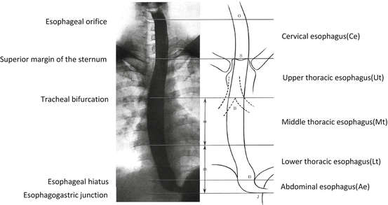
In the 4th edition (UICC, 1992) [4], together with the 3rd edition (AJCC, 1988) [10], the anatomical subsites of the esophagus were defined in the same fashion as in the 1st edition (JSED/JES, 1969) (Fig. 5.3) [15]. Consequently, all three stage classifications have the same definition for the esophageal subsites.
In the latest 7th edition (UICC, 2009) [7] however, the definition of the esophagogastric junction for adenocarcinoma was remarkably changed together with the latest 7th edition (AJCC, 2010) [14] according to Siewert’s Classification [29].
5.3.1.1 Definition of Esophagogastric Junction (C16.0) [7, 14]
Note: An adenocarcinoma with its epicenter within 5 cm of the esophagogastric junction and which extends into the esophagus is classified and staged using the esophageal scheme. Where the epicenter in the stomach is greater than 5 cm from the esophagogastric junction or is within 5 cm of the esophagogastric junction but without extension into the esophagus, then the adenocarcinoma is classified and staged using the gastric carcinoma scheme.
5.3.2 The AJCC Cancer Staging Manual
The anatomical subsites of the esophagus and the stomach in the 1st edition (AJC/AJCC, 1977) [8] were classified in the same fashion as the 3rd edition (UICC, 1978) [3]. Anatomical regions of the esophagus were defined by the distance from the upper incisor teeth.
In the 3rd edition (AJCC, 1988) [10], the anatomical subsites of the esophagus were labeled according to the ICD-O and defined in the same fashion as in the 1st edition (JSED/JES, 1969) (Fig. 5.3) [15].
In the 5th edition (AJCC, 1997) [12], the lower thoracic portion of the intrathoracic esophagus (C15.5) includes the intra-abdominal portion of the esophagus and the esophagogastric junction.
5.3.3 The Japanese Classification
The anatomical subsites of the esophagus—tumor location and anatomical esophageal nomenclature—were defined in the 1st edition (JSED/JES, 1969) (Fig. 5.3) [15]. In the 2nd edition (JSED/JES, 1972) [16], cancer of the esophagogastric junction was defined as a tumor limited between superiorly the lower and abdominal esophagus and inferiorly the upper third of the stomach, and lymph node groups for cancer of the esophagogastric junction (EC, E = C, CE)—N category—were classified.
In the latest 10th edition (JES, 2008) [25, 26], several criteria for clinical diagnosis of the esophagogastric junction are presented, and the zone of the esophagogastric junction is newly defined according to Nishi’s Classification [30].
5.3.3.1 Definition of the Esophagogastric Junction (EGJ) [25, 26]
The esophagogastric junction is on the border of the esophageal muscle and gastric muscle, and the location is clinically and pathologically diagnosed as
1.
The lower margin of the palisading small vessels in the lower esophagus by endoscopic findings
2.
Horizontally at the same level as the angle of His in an upper gastrointestinal series (UGI)
3.
Oral margin of the longitudinal fold of the great curvature of the stomach on endoscopic findings and UGI
4.
Obvious macroscopic caliber change in the resected esophagus and stomach
5.
The squamocolumnar junction (SCJ) is not always consistent with EGJ
5.3.3.2 Definition of the Zone of the Esophagogastric Junction [25, 26]
This zone is defined as within the region between 2 cm in esophagus and 2 cm in the stomach from the esophagogastric junction. The abdominal esophagus is included in this zone.
In the 1st to 11th editions of the General Rules for the Gastric Cancer Study (Japanese Research Society for Gastric Cancer, JRSGC), there was no description on the esophagogastric junction or of cancer in the esophagogastric junction. In the 12th edition (JRSGC, 1993) [31], and in the 1st English edition of the Japanese Classification of Gastric Carcinoma (JRSGC, 1995) [32], it is described that when a tumor was located in the upper third (C) and extended into the esophagus (E), then the case should be described as CE, and that this tumor in the esophagogastric junction should be described as CE or EC. Lymph node groups for dissection of regional lymph nodes—N categories—are added, when the tumor invades the esophagus.
5.4 T Category—Primary Tumor
5.4.1 The TNM Classification
In the 1st edition (UICC, 1968) [1], the T category was classified by the regional extension and morbidity. In the 2nd edition (UICC, 1974) [2], the T category was classified by the length of the tumor, circumferential extension, and extra-esophageal spread.
In the 3rd edition (UICC, 1978) [3], the TNM pretreatment clinical classification and the pTNM postsurgical histopathological classification were introduced. The latter was classified by the depth of tumor invasion in the same fashion as in the 2nd edition (JSED/JES, 1972) [16].
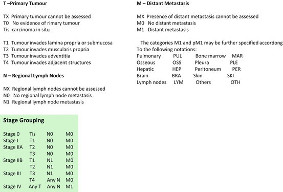
In the 4th edition (UICC, 1987) [4], the clinical T and pathological T categories were unified to the same classification as the pT category, as shown in Fig. 5.4.
In the latest 7th edition (UICC, 2009) [7], the T category is modified. T1 is divided into Tis, T1a (T1a-LPM and T1a-MM), and T1b and becomes the same as in the 9th edition (JSED/JES, 1999) [23, 24]. High-grade dysplasia is added into the same group as Tis. T4 is divided into two categories: T4a, where the tumor invades resectable organs, and T4b, where the tumor invades unresectable organs (Fig. 5.5).


Fig. 5.5
TNM categories and stage grouping in the seventh edition of the TNM Classification of Malignant Tumours (UICC, 2009) [7]
5.4.2 The AJCC Cancer Staging Manual
In the 1st edition (AJC/AJCC, 1977) [8], the same T category was adopted as that in the 2nd edition (UICC, 1974) [2]. In the 3rd edition (AJCC, 1988) [10], the T category was classified by the depth of tumor invasion in a similar way as in the 4th edition (UICC, 1987) (Fig. 5.4) [4]. After this unification, the T category of the AJCC Cancer Staging Manual becomes the same as that of the TNM Classification to date.
5.4.3 The Japanese Classification
In the 1st edition (JSED/JES, 1969) [15], the T category was classified by the extent of invasion to the adventitia: A0, no invasion to adventitia; A1, possible invasion to adventitia; A2, definite invasion to adventitia; and A3, invasion to neighboring structures. In the 2nd edition (JSED/JES, 1972) [16], clinical and histological T categories were introduced. The histological T category was classified by the depth of tumor invasion.
5.4.3.1 Histological T Categories [16]
ep: carcinoma in situ
mm: invasion to muscularis mucosa
sm: invasion to submucosa
mp: invasion to muscularis propria
a1: possible invasion to adventitia
a2: definite invasion to adventitia
a3: invasion to neighboring structures
In the 9th edition (JSED/JES, 1999) [23, 24], both clinical and histological T categories were unified and were classified only by the depth of tumor invasion.
Stay updated, free articles. Join our Telegram channel

Full access? Get Clinical Tree


