Modality
Clinical utility
Overall accuracy (%)
Computed tomography (chest, abdomen)
Invasion of local structures (airways, aorta)
≥90
Metastatic disease
≥90
Endoscopy
Local tumour (T) staging (operator dependent)
80–90
Endoscopic Ultrasonography (with or without fine-needle aspiration of lymph nodes)
Local nodal (N) staging (operator dependent)
70–90
Positron emission tomography
Metastatic disease, assessing response to neoadjuvant therapy
≥90
16.2.4 Staging
TNM staging is one of the most important and reliable prognostic variables. Standardised and accurate staging of cancer is important for uniform reporting and comparison of results from various centres. It also determines whether the intent of treatment is curative or palliative. It is based on clinical examination and information obtained by imaging: CT scan/PET–CT and/or endoscopic ultrasonography (EUS). The seventh edition of the AJCC TNM classification came into effect in 2009 [51].
Some of the key modifications from the sixth edition are:
1.
Inclusion of gastro-oesophageal junction tumours and tumours in the proximal 5 cm of the stomach extending into the oesophagus.
2.
T4 is subclassified as T4a (resectable cancer invasion) and T4b (unresectable cancer invasion).
3.
N staging is subclassified based on the number of positive regional lymph nodes (N1, 1–2 nodes; N2, 3–6 nodes; and N3, ≥7 nodes).
4.
M classification is redefined based on the presence of distant metastasis, and the term non-regional lymph node is eliminated.
5.
Histological grade and tumour location are incorporated.
6.
Separate stage grouping for adenocarcinoma and squamous carcinoma.
The new staging system has shown remarkable homogeneity within stage groups and excellent separation of survival curves between stages. The authors also welcome the separation of resectable (T4a) from unresectable (T4b) tumours. However, while the seventh edition is clearly superior in terms of prognostication, it is not ideal for baseline clinical staging or staging of patients who have undergone preoperative therapy. This is because the emphasis on nodal count rather than anatomic location and the introduction of histological grading make pre-resectional staging extremely difficult and highly likely to be inaccurate. Moreover, most of the data on which the stage grouping was based was drawn from western countries with a predominance of adenocarcinomas. Whether the same prognostic separation of the stage holds true for squamous oesophageal cancers remains to be seen.
16.2.5 The Tata Memorial Centre Experience
The authors’ institution, the Tata Memorial Centre, is the largest tertiary level cancer centre in the country and is a high-volume centre for the treatment of oesophageal cancer. Between 1,200 and 1,300 new patients with oesophageal cancer are seen every year, most of them presenting in advanced stage of disease or in an emaciated condition, precluding potentially curative treatment. Squamous oesophageal cancers predominate in a ratio of 80:20 and the most common location of tumours is in the lower third of the oesophagus. The typical diagnostic workup of patients with a good performance status includes a detailed flexible fibre-optic upper gastrointestinal endoscopy with mapping of the disease and biopsy, PET–CT scan with contrast, pulmonary function tests with diffusion coefficient of carbon monoxide (DLCO) and cardiac evaluation. Flexible fibre-optic bronchoscopy is performed in patients with upper and middle third lesions and those with an obvious change of voice; endoscopic ultrasonography is done selectively for patients with low-volume disease on CECT scan (to confirm early disease amenable for upfront surgery) or in borderline resectable disease after neoadjuvant therapy. This diagnostic workup is curtailed in patients who are emaciated and not fit for radical therapy and in patients with obviously metastatic disease. Patients who are high risk for surgery due to co-existing co-morbidities undergo a thorough cardiopulmonary evaluation and are discussed in a special “high-risk multidisciplinary team” meeting by surgeons, intensivists and critical care specialists, anaesthesiologists and pulmonary physicians to optimize them prior to surgery. The preferred therapeutic approach is discussed in a subsequent section of the chapter.
16.3 Treatment of Squamous Oesophageal Cancer in India
16.3.1 Treatment
India is a vast and populous country with significant resource constraints. The wide variation in the availability of facilities and technical expertise across different regions has made standardization of treatment a difficult process. While the establishment of 27 regional cancer centres across the country has partially addressed the issue, the urban–rural divide and between-centre variability of care are still considerable. Efforts by the authors’ institute and the Indian Council of Medical Research (ICMR) have culminated in the establishment of uniform oesophageal cancer treatment guidelines tailored to the country’s varied levels of expertise and availability of infrastructure. One of the core recommendations of the guidelines is the establishment of multidisciplinary teams for the management of oesophageal cancer. While some major cancer centres in India have a multidisciplinary team including a surgical, medical and radiation oncologist in place, several others do not, and one of the biggest challenges has been to ensure the same standards of care and decision-making regardless of whether the patient initially presents to a surgeon, gastroenterologist and medical or radiation oncologist.
16.3.1.1 Patient Evaluation
The initial evaluation of the patient includes the assessment of physical (ECOG performance) status, oral hygiene, nutrition and cardiopulmonary status. This is particularly important in the Indian scenario, where patients generally present in an advanced stage and in poor general health. Generally, only patients who are ECOG performance score (PS) 0 or 1 are selected for radical treatment. Assessment of oral hygiene is necessary because of the high prevalence of tobacco chewing in India [27, 29] and the possibility of co-existing oropharyngeal malignancy. Since most patients present with significant dysphagia and some degree of nutritional impairment, assessment of nutritional status and early institution of rehabilitation is key. The enteral route is the preferred route of nutritional rehabilitation due to its inherent advantages of keeping the gut in use, as well as the ease of administration and relatively low complication rate compared to parenteral nutrition [52]. All patients considered for radical treatment undergo extensive evaluation of cardiopulmonary status including pulmonary function tests (PFT), 2D echocardiography and, in select cases, stress cardiac testing. Pulmonary rehabilitation is started at the outset for all patients planned for radical treatment with the active involvement of the chest physician and physiotherapists. Early institution of chest physiotherapy and tobacco and alcohol cessation are routinely advocated as soon as a diagnosis of oesophageal cancer is made.
16.3.1.2 Principles of Management
Broadly, decisions regarding the treatment are based on the anatomical location and stage of disease and the performance status of the patient. The authors’ repeated emphasis on the performance status of the patient is primarily because poor general health precludes potentially curative treatment in considerable proportion of patients in India. Concurrent radical chemoradiation is the preferred therapeutic strategy for lesions in the upper third of the oesophagus, i.e., within 5 cm of the cricopharynx, while surgery is the preferred treatment for lesions in the middle and lower third oesophagus. Early stage lesions (T1/T2, N0) are usually treated by surgery alone for middle and lower third lesions. Endoscopic mucosal resection (EMR), though a less morbid procedure, is not widely practised in India primarily due to the fact that very few patients present at a stage amenable to the procedure and also due to the limited availability of expertise in select centres across the country. Patients with locally advanced disease (T3/T4, N+) undergo multimodality treatment, generally with neoadjuvant chemotherapy [53, 54] or neoadjuvant chemoradiotherapy followed by surgery. Patients with metastatic disease are usually treated with palliative radiotherapy or oesophageal stenting or a combination of the two and rarely with palliative chemotherapy.
16.3.2 Surgery
Surgery is the preferred modality of treatment for middle and lower third oesophageal cancer [55–58]. Most patients in India with early stage disease (T1/T2, N0) are considered for upfront surgery while patients with locally advanced disease undergo surgery after neoadjuvant therapy. Rarely, patients with residual disease after radical chemoradiotherapy are taken up for oesophagectomy albeit at the cost of significantly higher post-operative morbidity. In spite of the established role of surgery in the radical treatment of oesophageal cancer, there is very little consensus on what constitutes a standard oesophagectomy in terms of approach, extent and template for lymph node dissection. This may, in part, be because there is no organ-specific surgical training program in India. Oesophageal resections in India are performed by surgeons from varied surgical specialties including general surgery [55, 56], gastrointestinal surgery [57], thoracic surgery [58] and surgical oncology [59].
16.3.2.1 Approach
Transthoracic oesophagectomy predominantly by a modified McKeown three-stage procedure is considered to be the standard approach by most thoracic surgeons and surgical oncologists while most general and gastrointestinal surgeons prefer a transhiatal approach particularly for lower third tumours [55–59]. In a large series of 367 transhiatal oesophagectomies performed over a period of 18 years at the All India Institute of Medical Sciences, the 5-year overall survival was 38 % with a post-operative mortality rate of 12 %. Since there is no strong evidence favouring one approach over the other, both approaches are widely practised in India with a bias towards transthoracic approach in high-volume oncology centres. In these centres, transhiatal resection is performed in limited numbers as a compromise surgery in patients with poor pulmonary function or extensive pulmonary fibrosis precluding transthoracic resection.
16.3.2.2 Lymphadenectomy
Lymphadenectomy for oesophageal cancer is a controversial topic in India, as in many other parts of the world [60]. Surgical oncologists who predominantly perform transthoracic oesophagectomies place more emphasis on extensive lymph nodal clearance. Infracarinal nodal dissection or a standard two-field dissection is considered to be the standard template for dissection by most surgeons performing a transthoracic oesophagectomy. In India, very few centres with high volumes of oesophageal surgery practice three-field lymphadenectomy routinely. The increase in lymph node yield with more radical lymphadenectomy needs to be balanced against an increased post-operative morbidity, primarily with recurrent laryngeal paresis and pulmonary complications. In contrast, the lymph node yield achieved by a transhiatal resection is low and is usually limited to the perioesophageal lymph nodes. However, as mentioned in the previous section, transhiatal resections are usually performed only as a compromise surgery in high-volume centres.
16.3.2.3 Minimally Invasive Surgery
Surgeons in India were early to adopt minimally invasive oesophagectomy. A few high-volume centres have published data showing better results with a minimally invasive approach with respect to pulmonary morbidity and operative blood loss [57, 59, 61–63]. A prospective study comparing minimally invasive oesophagectomy with open oesophagectomy [63] demonstrated comparable results in terms of lymph node yield (9.5 vs 7.3), duration of surgery (312 vs 262 min), average blood loss (276 vs 313 mL) and morbidity (26.5 vs 28.6 %). A larger series [57] of 463 thoracoscopic oesophagectomies demonstrated a lower morbidity rate (16 %) and post-operative mortality rate (0.9 %). However, no long-term (survival) outcome data is available from any of these studies. Different surgical groups in India use different patient positions for thoracoscopic oesophagectomy with lateral, prone and, more recently, semi-prone positions being utilized based on surgeon preference. The prone or semi-prone position offers the advantage of not requiring lung isolation for thoracoscopy, whereas the lateral position offers better exposure to the superior mediastinum for radical lymph node dissection. The authors’ preference is to perform MIS oesophagectomy through the lateral approach. Robotic surgery for oesophageal cancer has just started in India and is confined to few centres currently. A series of 32 robotic oesophagectomies [64] showed comparable results to thoracoscopic oesophagectomy. However, no distinct advantage over thoracoscopic oesophagectomy has been demonstrated.
16.3.2.4 Reconstruction
The stomach is the preferred conduit for reconstruction, and in cases where the stomach is not available, the colon, either the right or left side, is the preferred alternative. The posterior mediastinum is the most commonly used route of reconstruction, the retrosternal route being used only when the patient is being considered for post-operative radiotherapy to the mediastinum or when the surgeon adopts an abdomen-first approach to a transthoracic oesophagectomy. A small randomized study of 49 patients comparing posterior mediastinal versus retrosternal conduit placement [65] found both routes to have comparable outcomes. The anastomosis is usually performed in the neck either by a stapled or handsewn technique [66]. Both techniques are widely practised in India depending upon surgeon preference and cost constraints. Some clinical trials on anastomotic technique are described in a subsequent section of the chapter.
16.3.3 Multimodality Management
India was late to embrace multimodality management in oesophageal cancer. This may have been primarily because of the delayed establishment of multidisciplinary teams and also the fear that multiple modalities of treatment may not be well tolerated by the generally frailer Indian patients. In view of the strength of evidence supporting neoadjuvant therapy currently, patients with locally advanced potentially operable oesophageal cancer are treated with either neoadjuvant chemotherapy [53, 54] or neoadjuvant chemoradiotherapy. The common chemotherapy regimens include doublets consisting of cisplatin with 5-fluorouracil or cisplatin with paclitaxel, while few centres use triplets of cisplatin, 5-fluorouracil and either paclitaxel or docetaxel, which have superior response rates at the cost of higher morbidity. The commonly followed schedule is to administer three cycles at three-weekly intervals followed by reassessment with CT scan imaging and surgery between 4 and 6 weeks after the last cycle of chemotherapy. The results with neoadjuvant chemotherapy have been encouraging in terms of tolerability and completion of planned treatment; however, no long-term outcome data is available. Neoadjuvant chemoradiotherapy is also rapidly gaining popularity in India. The most commonly used protocol is the CROSS protocol, i.e., radiation 41.4 Gy in 23 fractions of 1.8 Gy over 5 weeks with concurrent weekly chemotherapy, paclitaxel 50 mg/m2 and carboplatin at AUC 2. Most centres are stringent in patient selection for this regimen, and the early results have been very encouraging.
Post-operative radiotherapy or chemoradiotherapy is not practised as a routine after oesophagectomy. The use of adjuvant radiotherapy is restricted to patients with positive resection margins and, occasionally, patients with significant residual metastatic lymphadenopathy after neoadjuvant chemotherapy.
16.3.4 Chemoradiotherapy
Chemoradiotherapy is the primary modality of treatment of upper third oesophageal cancers and locally advanced middle and lower third cancers that are unresectable. It is also the treatment of choice in patients who are medically inoperable or unwilling to undergo surgery. The most widely practised and well-tolerated regimen includes radiotherapy to 66 Gy in 33# in 6.5 weeks with concurrent weekly cisplatin 35 mg/m2, 5–6 cycles [67]. In institutes with facilities for intraluminal brachytherapy the radiation regimen may be changed to teletherapy 50 Gy in 25# in 5 weeks followed by 2# of high-dose rate intraluminal brachytherapy of 12 Gy after 2 weeks, the chemotherapy regimen remaining the same. Several concurrent chemotherapy regimens are practised including three-weekly cisplatin and 5-fluorouracil and three-weekly paclitaxel and cisplatin along with standard doses of radiation.
16.3.5 Palliative Therapy
The emphasis of management in patients presenting with metastatic oesophageal cancer is on early palliation of dysphagia. Patients with metastatic disease but grade 3 or less dysphagia are treated with palliative radiotherapy with or without stenting [68]. Patients with absolute dysphagia who need immediate palliation are treated with oesophageal stents, most commonly self-expanding metal stents [69]. A few centres offer intraluminal radiotherapy for metastatic and locally advanced oesophageal cancer and have been found to offer faster and sustained palliation of dysphagia [70]. Rarely patients with bulky disease obstructing the tracheobronchial tree as well as the oesophagus are treated with double stents i.e., tracheal and oesophageal stents.
16.3.6 The Tata Memorial Centre Experience
At the authors’ institute, patients with early (T1 or T2 with N0) disease are treated with primary surgery, while those with more advanced (T3 or T4a or N+) disease are treated with neoadjuvant chemotherapy (NACT) or chemoradiotherapy (NACTRT) followed by surgical resection. While the default option is NACT for most patients, eligible patients are currently getting randomized in a phase II trial comparing the two strategies. The diagnostic workup and treatment guidelines are summarized in Fig. 16.1. Over 1,700 surgeries have been performed for oesophageal cancer over the last 10 years. The preferred choice of surgery is a transthoracic three-stage oesophagectomy while transhiatal oesophagectomy is occasionally performed as a compromise procedure in patients with borderline fitness or extensive pulmonary fibrosis. Elective three-field lymphadenectomy is done in all patients with supracarinal disease and those with radiologically or metabolically metastatic supracarinal lymphadenopathy. Patients without these features are considered for randomization to a trial comparing standard two-field with elective radical three-field lymphadenectomy. Minimally invasive oesophagectomy (thoracoscopy and/or laparoscopy) is performed in approximately half of the patients undergoing transthoracic oesophagectomy. The preferred conduit is the stomach and the posterior mediastinum, the most common route of reconstruction. Oesophagogastric anastomosis is performed in the neck by a triangulated stapled anastomosis. A nasojejunal tube is placed intraoperatively for post-operative enteral feeding.
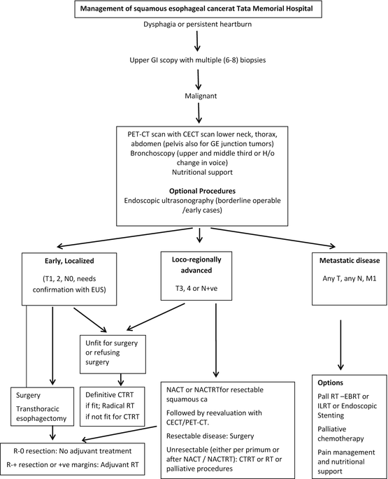

Fig. 16.1
OSCC treatment algorithm at the Tata Memorial Hospital
Preoperative preparation includes chest physiotherapy, incentive spirometry and nutritional rehabilitation along with smoking cessation. Anti deep vein thrombosis (DVT) prophylaxis is started 12 h prior to surgery and continued post-operatively. Prophylactic antibiotics are given preoperatively and repeated once after 3 h intraoperatively and are not continued routinely in the post-operative period. Most patients are extubated immediately post-operatively on table and shifted to a recovery ward rather than the intensive care unit. Physiotherapy and active mobilization are started soon after shifting to the recovery ward. Enteral (nasojejunal) feeding is started the morning after surgery and stepped up gradually to full enteral feeds by the evening of the second post-operative day. The nasogastric tube is clamped on the second post-operative day and removed by the same evening if the chest radiograph shows no gastric tube dilatation. Routine laryngoscopy examination is done to check the vocal cord status on the fifth post-operative day and oral liquids started on the sixth post-operative day. Contrast swallows are not done prior to starting orals and patients are on full solid feeds by the 8th post-operative day. Uncomplicated patients are discharged by the tenth post-operative day. The post-operative major morbidity and mortality are 19.9 and 5.9 %, respectively. Common post-operative complications include pulmonary complications (27.1 %), anastomotic leaks (8.8 %), vocal cord paresis (31.4 %, of which 6.3 % have permanent palsy) and thoracic duct injuries (1.3 %). The 5-year survival of patients undergoing total oesophagectomy was 42 % with a median survival of 36 months (95 % confidence interval, 25.5–46.5 months).
16.4 Research in Oesophageal Cancer in India
Research on oesophageal cancer in India has a long history. The main areas of focus in oesophageal cancer research have been the possible aetiological factors and associations with squamous oesophageal cancer, the choice of primary treatment for the disease, modifications in surgical technique, the role of neoadjuvant and adjuvant treatment and palliative treatment options.
16.4.1 Epidemiology Research
Epidemiological research from the Kashmir Valley, which is a high incidence area for squamous oesophageal cancer, established that low socio-economic status was an independent risk factor [15]. A large case–control study, matched for age, sex and geographic area, showed a strong inverse association between higher education and wealth status and OSCC risk. The same study also established the probable aetiological role of “hookah” smoking and “nass chewing” on oesophageal squamous cell cancer with odds ratios of 1.85 and 2.88, respectively [17]. In a small study evaluating the prevalence of human papillomavirus (HPV) strains in OSCC, researchers found that a high proportion (87 %) of patients with OSCC harboured high-risk HPV strains [37]. While association between HPV strains and OSCC is already established and the study supported the hypothesis of persistent oncogenic viruses in cancer development, a larger study would be required to firmly establish causation. In a study of epigenetic, genetic and environmental interactions in OSCC, significantly higher methylation frequencies were noted in tobacco chewers compared to non-tobacco users for all the four genes (p16, DAPK, BRCA1 and GSTP1) studied [33]. Betel quid chewing, alcohol consumption and a null GSTT1 genotype had maximum risk for OSCC without promoter hypermethylation whereas tobacco chewing, smoking and null GSTT1 variants were found to be associated with OSCC with promoter hypermethylation on logistic regression analysis [33].
16.4.2 Primary Treatment
One of the two randomized trials [71, 72] comparing surgery with radical radiotherapy for localized oesophageal cancer was conducted in the authors’ institute. Although this trial was primarily designed to evaluate quality of life in patients treated with surgery or radiotherapy, it established that surgery was far superior to radiotherapy even for overall survival [72]. The study randomized 99 patients to either surgery alone (n = 47) or radiotherapy alone (n = 52). Outcomes with respect to disease-specific symptoms, which was the primary outcome, were consistently superior in the surgery arm; specifically, the quality of swallowing, which is an important endpoint of treatment of oesophageal cancer, was superior in the surgery arm compared to the radiotherapy arm. The secondary endpoint of survival was vastly superior in the surgery arm compared to the radiotherapy arm (p = 0.002) [72]. To date, this is one of only two randomized trials [71, 72] performed so far to address this important question.
Stay updated, free articles. Join our Telegram channel

Full access? Get Clinical Tree


