Jay A. Yelon and Fred A. Luchette (eds.)Geriatric Trauma and Critical Care201410.1007/978-1-4614-8501-8_5
© Springer Science+Business Media New York 2014
5. Skin, Soft Tissue, and Wound Healing in the Elderly
(1)
Department of Surgery, University of California, Davis Medical Center, Shriners Hospitals for Children Northern California, 2425 Stockton Blvd., Sacramento, CA 95817, USA
Abstract
Aging leads to significant changes that predispose people to problems with their skin. Aging-related alterations in skin can be classified into “intrinsic” changes that are simply related to the natural aging processes and “extrinsic” effects that are due to exposure to the ultraviolet rays of the sun. The elderly tend to have a thinner dermis, flattening of the dermal-epidermal junction, and decreases in the dermal appendages. These changes lead to an increased propensity of skin to tear and shear. The decreased density of appendages impairs the ability to reepithelialize superficial wounds. As everyone knows, the skin also tends to stretch (or sag) which allows for contraction of wounds with less functional impairments. There are also body composition changes to the muscle and fat stores that decrease padding that increase the risk for pressure damage. Age-related disease processes such as urinary incontinence, diabetes mellitus, vascular disease, malignancy and its treatment, and other neurologic diseases predispose patients to increased risk. As people age, their reaction times may decrease to prevent injury, and confusion may lead to increased risky behavior. The incidence of pressure ulcers, arterial ulcers, venous stasis ulcers, and diabetic ulcers increases with age. This chapter will cover the pathophysiology of aging of the skin, the risks that increase the risk for wounds in the elderly, chronic wounds, prevention of wounds, and the outcomes of the elderly with wounds.
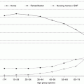
In the industrialize world the birth rate is down, and with modern healthcare people are living longer so that the elderly are occupying a much larger percentage of the population. US census results reveal that in 2010, 24 % of the population was greater than 55 years of age, and 13 % was greater than 65 years of age. These values are projected to be 31.1 % >55 years, 20.2 % >65 years, and 4.3 % >85 years in 2050 [1]. All practitioners are going to be exposed to more geriatric patients, so familiarity with skin problems will become essential knowledge. Not only are the elderly at greater risk for skin breakdown but they also have skin changes that alter their ability to heal. Many treatments for diseases in the elderly (steroids, chemotherapy, and radiation) impair tissue repair. Chronic wounds – diabetic, vascular, venous stasis, and pressure ulcers – are much more common in the geriatric population. Chronic wounds are a huge economic burden for today’s healthcare and for the individual; they frequently accompany the person for the rest of his or her life. Pressure ulcers are considered a “never event” by governmental health agencies that, if they occur, may lead to loss of reimbursement. Since simple injuries often lead to major wounds that fail to heal, prevention efforts are essential to reduce the burden of chronic wounds in the elderly. While there are increased problems with healing in the elderly, their wounds can be treated and lead to successful outcomes. This chapter will review the factors that increase the risks for wounds in the elderly, describe the pathophysiology of chronic wounds, and discuss prevention and describe strategies for treating those wounds.
Skin Changes with Aging
The skin changes related to aging are well documented in the dermatology literature [2–7]. Typically, skin alterations due to aging are classified into “intrinsic” and “extrinsic” changes. Intrinsic changes are those that occur “within the body” as part of the normal aging process and are independent of environmental exposure. Extrinsic changes are those alterations that are induced by environmental forces – most notably the ultraviolet portion of the sunlight. While it is difficult to differentiate which factors are totally intrinsic versus extrinsic, it is clear that extrinsic factors accelerate the degenerative changes in the skin. Everyone is aware of the changes that occur in skin that is abused by sun exposure or just poor self-care. The skin becomes thinner, dryer, more wrinkled, sags, and has variable pigment changes. Clearly, sun exposure increases the risks of skin cancers of all types. While these changes will occur with aging, good skin care, especially protection from the sun, will slow these changes.
The structural changes of skin that occur with aging are well documented. The epidermis tends to become thinner with aging but thickens in response to ultraviolet light damage. The dermal-epidermal junction becomes flatter. The flattening of the normal rete pegs of the dermal-epidermal junction weakens the resistance to epithelial shear. In other words, the elderly are more prone to superficial wounds from minor shear forces. There are also significant changes to the normal skin adnexa – hair follicles, oil glands, sebaceous glands, and other dermal appendages. Sebaceous glands are decreased which leads to more dryness of the skin. In addition, there is decreased replacement of lipids in the stratum corneum which interferes with the normal barrier function of the epidermis. Hair follicles clearly change in many parts of the body. They heal more slowly and change in distribution. Clearly, male and female alopecia (baldness) is the most recognizable hair change, but hair follicles increase in size and decrease in density [6].
The significance of the dermal appendage changes is that healing of superficial wounds is impaired. To heal a partial-thickness wound (such as a superficial burn or blister), the dermal appendages are required. Normally, reepithelialization takes place in two areas of the skin, the edge of normal skin and from dermal appendages [8]. At the edge of the wound, the basal cells of the bottom layer of the epidermis are stimulated to migrate across the wound by three factors: loss of cell-cell contact, stimulation by growth factors (epidermal growth factor, transforming growth factor-α, and keratinocyte growth factors 1 and 2), and contact with proteins of the exposed wound (fibronectins, collagen type 1). Migration from the original wound edge stops after around 1–2 cm in a full-thickness wound, and the remainder of the closure is by contraction. In a superficial wound, epithelial migration also occurs in the epithelial cells of the dermal appendages, especially hair follicles. The higher the hair follicle density in a wound, the faster it can reepithelialize. As an example, if a hair-bearing scalp is used as a split-thickness skin graft donor site, it will heal within 4–5 days. If the skin adnexa are sparse in number, such as occurs in a lower leg with impaired circulation, then healing may take weeks. Thus, the problem with the decreased density of dermal appendages in the elderly is that their ability to reepithelialize a wound is impaired. I have observed very superficial wounds in a hairless, elderly patient that never reepithelialize and thus are said to “convert” to full-thickness. They did not “convert” but instead had no ability to reepithelialize.
There are significant dermal changes in the skin due to aging. The dermis is also the main target of ultraviolet light damage [2]. With increasing age there is a decrease in dermal cells (macrophages, fibroblasts, mast cells) and a decrease in antigen-presenting cells (Langerhans cells) which results in a decrease in the immune function. The dermis becomes thinner and the collagen becomes less organized. The collagen molecules actually become larger but have more fragmentation and less orientation along lines of stress. The activation of matrix metalloproteinases by ultraviolet light exposure may contribute to these collagen molecule changes. This degradation in collagen is reflected in studies which demonstrate that the tensile strength of skin is decreased with age [9]. The dermal hydration decreases, and while the amount of elastin changes little, it becomes more fragmented. Thus, skin becomes less elastic, more wrinkled, and more prone to tearing or lacerations. Everyone knows that with aging comes looser skin that sags with gravity. While looser skin is considered undesirable in our society, it does benefit the healing of the elderly with small full-thickness wounds. These wounds heal by contraction so that the loose skin tends to not interfere with this process as it would in a younger person. Tension tends to interfere with wound contraction, so the process may be augmented with looser skin. Clearly, contraction can lead to contractures if it occurs over functional areas such as joints. Since looser skin allows for closure with less tension, the elderly have a lower risk for contracture. In other words, allowing a wound to contract may be an alternative to surgical repair.
Besides the actual structural changes to the skin, there is a generalized decrease in sensation, decreased vascularity, and impaired lymph flow with aging. The sensory changes occur in a distal to proximal fashion and are especially related to decreased cold/warm sensory abilities [10]. There are actual decreases in the density of thermal sensory receptors, and some suggest that there is a decrease in peripheral nerve density. Studies suggest that there is altered angiogenesis associated with decreased cutaneous vascular reactivity [2–7]. In response to sun damage, there may be an increased angiogenic response, but the new vessels are more disorganized and more prone to leaking proteins. With impaired lymph flow there is an increase in edema – which impairs healing. In addition, impaired lymphatic function decreases the ability to fight infections or contract wounds. There are obvious pigment changes that occur in skin with aging. Melanocytes decrease in numbers, but with ultraviolet light exposure, there are more spotty areas of hyperpigmentation. Many cells develop mutations that alter local areas leading to “age spots” such as keratoses and nevi. While not covered in this review, skin cancers increase with aging. All of these changes are accelerated with sun exposure. In addition, increased bruisability from the use of antiplatelet drugs or anticoagulants will often lead to pigment changes from the retained heme products.
Finally, changes beneath the skin contribute to the risk of skin injury in the elderly. There is a tendency to lose muscle mass from either decreased exercise or activity. Fat stores are often (but not always) reduced with aging. These changes are clearly accelerated with malnutrition – a factor that impairs wound repair. The significance of these changes is that the loss of padding tends to expose bony prominences to increased pressure and chronic breakdown. Incontinence of urine or stool increases the risk for maceration which increases the risk of shear injury. As people age, they become slower in their ability to respond to a dangerous situation. Their reflexes tend be slower so that they have more difficulty escaping an injury. I have observed many elderly patients who were unable to extinguish flaming clothing or escape scalding water. This slowing of reflexes and an impaired ability to respond to injury lead to more extensive and deeper wounds. We know from burn studies that the elderly have a much lower ability to tolerate large wounds, so a small injury can be fatal. Finally, people at the extremes of ages become more dependent on others for care. An unfortunate consequence of this unwanted dependency is that there is an increase in the risk for elderly abuse. One must always be wary if an injury does not fit the “story” of how it occurred. Just as for children, caregivers are obligated to report suspected abuse.
Diseases of the Elderly Affecting Wound Healing
Fortunately, healing processes remain fairly normal in healthy people until the extremes of ages. Since many elderly take good care of themselves, they tolerate surgery and minor injuries quite well. It is important to recognize, however, the factors or diseases that impair tissue repair. These inhibitors of wound healing affect all age groups, but they are, unfortunately, more common in the elderly population. One must be aware of these factors that may delay or prevent healing if he or she is planning surgery or treating a wound.
Malnutrition
Studies have shown for over 100 years that malnutrition impairs wound healing [11–14]. The impairment exists with total protein/calorie (marasmus) malnutrition or with protein (kwashiorkor) malnutrition. Simply, if one holds nutritional support at the time of wounding, a marked impairment in tensile strength will result within 1–2 weeks [15]. The clinical significance of malnutrition relates to the risks of complications with surgery such that if a patient has lost weight from a malignancy or from an inability to eat, then there is a much higher risk for dehiscence. The hidden side of this healing impairment is that altered healing could lead to a bowel anastomosis leak which in turn, may lead to an abscess. Since the metabolic reserve is reduced in the elderly, this complication of failing to heal often leads to sepsis, multiple organ dysfunction syndrome, and ultimately death. The clinician can reduce complications by assessing the nutritional status of the elderly and providing supplemental nutrition prior to surgery. In addition, an aggressive approach to perioperative nutrition may make the difference between normal healing without complications and a rocky course and ultimate death.
There are some vitamins and micronutrients that influence tissue repair. Vitamin C is essential for the hydroxylation of proline or lysine in the formation of normal procollagen triple helices [16]. When there is a deficiency, collagen is not properly produced, and people may suffer from scurvy. Since there is a balance between collagen formation and breakdown during the maturation phase of scar formation, recently healed wounds may break down. In addition, vitamin A has been found to augment tissue repair [17, 18]. Several minerals such as zinc [19, 20] and copper [20] are essential for normal healing, and when they are deficient, problems may result. We have noted that patients with subnormal copper levels have impaired healing of their burn wounds [21]. A deficiency in arginine may also lead to altered tissue repair [22].
Diabetes Mellitus
Diabetes mellitus is a major cause of healing problems in people of all ages [23–25]. Since the disease is seen more commonly in the elderly, it is important to know its impact on tissue repair. Some statistics are important to emphasize its impact on the development of chronic wounds. Twenty percent of hospital admissions in diabetes are related to wound healing problems. Twenty-five percent of diabetic patients will have a foot ulcer during their lifetimes, and 50 % of all nontraumatic amputations are related to diabetes mellitus. A common scenario is that a diabetic patient will not notice a pebble in their shoe due to their impaired sensation. This will create a small wound that goes unnoticed, and eventually a wound is noticed and not properly treated. When the patient finally seeks care, the wound is infected with purulence tracking up the fascial planes of the foot. This patient often presents with cellulitis or invasive infection that leads to an amputation.
Even if a wound is detected early, tissue repair is significantly impaired. As an example, Margolis reviewed several clinical trials that tested treatments for diabetic ulcers [26]. He collected the “controls” which received “standard” treatment. What he found was that only 31 % healed within 20 weeks of aggressive therapy. It is likely that the two-thirds that did not heal stayed open for the remainder of their lives. There are several reasons why tissue repair is impaired in patients with diabetes mellitus [23–25]. First of all, peripheral vascular disease is increased in this population. Diabetics not only suffer from macrovascular disease but also from microvascular disease where there is thickening of the capillary basement membrane. This microvascular disease leads to impaired delivery of oxygen and nutrients from an increase in edema which impairs diffusion. This process also impairs leukocyte migration into the wound. If the patient suffers from renal disease, healing is inhibited further by uremia and its resulting edema [27].
The second factor that contributes to alter healing in diabetes mellitus is its tendency to cause peripheral neuropathy. Loss of sensation progresses from distal to proximal in the extremity so that the feet are usually involved first. As stated earlier, people with neuropathic feet do not sense injury, and thus minor injuries tend to worsen or go unrecognized by the patient. We have recently reviewed our 10-year experience with diabetics who burn their feet [28]. It is common for them to walk outside on hot pavement or try to warm their “cold” feet with hot water or by placing them near space heaters. We admitted 68 patients with thermal injury to their feet during that period, and the incidence is increasing. As for other types of diabetic wounds, the patients admitted for burns to their feet tended to have prolonged hospital stays and frequent graft failure. Another often unrecognized consequence of neuropathy is that the foot loses its normal feedback to maintain the arch. The foot thus tends to flatten which leads to increased pressure on the first or second metatarsal head. The classic diabetic foot ulcer is a wound that is on the plantar surface on the first or second metatarsal head. The final consequence of neuropathy is that with loss of normal sympathetic innervation, the skin loses its ability to sweat and thus tends to dry and crack. These cracks may then be a site for infection.
There is an increased risk for infection for wounds that form in the diabetic patient. It is well known that hyperglycemia leads to an impaired ability to fight local infection. There are also studies that suggest that leukocyte migration and function is impaired. The impaired ability to fight infections predisposes diabetics to a higher risk for amputation. In addition, there are several metabolic factors that may contribute to impair healing that are covered in other reviews [25, 29]. One interesting concept is that hyperglycemia may lead to deposition of glucose by-products known as advanced glycosylation end-products (AGES) in the tissues. There are “receptors for advanced glycosylation end-products” (RAGES) that detect these products of hyperglycemia and stimulate an inflammatory response. One theory is that activation of RAGES may lead to the chronic inflammatory state (“metabolic syndrome”) of diabetes mellitus and obesity [30]. This chronic inflammatory state may also contribute to impaired tissue repair.
Since healing is so impaired in diabetic patients, it is essential that prevention efforts are made to prevent a wound from developing. The Wound Healing Society has published guidelines for the treatment and prevention of diabetic wounds [31, 32]. All clinicians who treat diabetes mellitus should discuss the risks of foot ulcers with their patients. Diabetics should inspect their feet daily and be extremely careful with the care of their nails. Podiatrists are extremely helpful in these matters. Diabetics with neuropathy should always wear well-fitted shoes and be especially vigilant after the first few days of wearing new shoes. Any new wound should be treated aggressively and early. People with diabetes mellitus should always avoid walking outside while barefoot and never warm their insensate feet with any heated agent. Once a wound develops, they should be treated with something that “off-loads” the pressure point on the wound (metatarsal head). “Total contact casts” have been found to be effective [33]. Studies suggest that topical growth factors or skin substitutes may be effective, but they are extremely costly [34–36]. Vascular disease should be treated if present. Unfortunately, our success with treating these wounds is only marginal, so prevention is essential.
Therapies That Alter Wound Healing
Wound healing involves the recruitment and rapid proliferation of many different cell types. It makes sense, then, that any drug that impairs rapid proliferation of cells alters tissue repair. Unfortunately, the strategy for dealing with many diseases includes suppressing the inflammatory response, which also requires rapid proliferation of cells. Steroids have been known for decades to impair tissue repair, and their use should be minimized if possible to allow for better healing [37, 38]. The treatment of cancer also involves the killing of rapidly proliferating malignant cells, so it is also obvious that chemotherapy agents [39, 40] or radiation [41, 42] impairs tissue repair. With the advent of neoadjuvant therapy (chemotherapy and/or radiation) combined with surgery, it is clear that one must be extremely careful with the healing of these patients. One must optimize their nutritional status if they are to undergo these combined treatments. There are very few agents that augment healing in these situations, but vitamin A has been shown to at least partially reverse impairments due to steroids or radiation [17, 18]. Growth factors may also play a role in improving healing, but these are based on animal studies [43–49].
Neurologic Diseases
Neurologic diseases do not impair wound healing, but they do predispose the elderly to the risk of developing wounds. Dementia leads to forgetfulness and risky behavior that may lead to injury. The person may forget to turn off stoves or fail to practice safe techniques for self-care. People with dementia have a more difficult time with cleanliness and maintaining a diet and thus may not clean wounds and tend to be more malnourished. Neuropathies have previously been mentioned as a risk for many types of wounds. Any loss of sensation clearly predisposes a person to pressure sores since pain is the main warning sign of chronic pressure. Tremors may predispose the elderly to spills and an inability to quickly react to a dangerous situation. Incontinence may lead to maceration of the skin which in turn increases the risks for abrasions or tears with moving. Seizures are risky in people who cook or are around hot items since during the seizure, they will not react to an injury. People who seize while cooking or bathing frequently sustain very deep burns.
Problem Wounds of the Elderly
There are specific types of wounds that all practitioners must know about when treating the elderly. These wounds are relatively easy to prevent but are particularly difficult to treat once they are present. These chronic wounds are a significant burden to society in cost and interference with normal living. They may occur in any age group, but they are more common in the elderly. Since they are a major contributor to morbidity in the elderly, one must know how to diagnose and treat these problem wounds. The Wound Healing Society has recently published consensus guidelines in the prevention and treatment of these problem wounds which provide hundreds of references [50–55].
Pressure Ulcers
Pressure ulcers may develop at any age, but they are commonly manifested in the elderly as they develop reduced ability to move or after the development of neuropathy [50, 51, 56]. These wounds are found in around 10 % of inpatients. The pathophysiology is simple; any pressure on the skin and underlying tissues of greater than 30 mmHg that persists for a prolonged period of time can lead to enough ischemia to create a pressure ulcer. Normally, pressure produces pain which leads to a shift in the body to redistribute the pressure to another area. When we sleep we are constantly and subconsciously moving. Even intoxicated people will move to prevent these wounds. When people lose sensation, such as after paralysis, or when so ill that they are unable to move (such as in an intensive care unit), they are prone to pressure ulcers. Nurses play a major prevention role by repositioning patients from side to side. Not uncommonly, however, pressure ulcers develop where bony prominences create pressure. The classic sites are in the presacral region, the occiput, and on the heels. There are scoring systems that grade the severity of pressure ulcers that are useful for documentation. Pressure ulcers are staged as 1 when the skin is intact but just reddened for a prolonged period, 2 when the skin has been broken, 3 when the ulcer has become full-thickness, and 4 when the wound involves deeper tissues [56, 57]. The wound management becomes more difficult with increasing severity. The usefulness of this paradigm, however, has been recently challenged [58]. It is unclear of the relevance of a stage 1 ulcer, and the pathophysiology varies between stage 2 and stage 3 or 4 ulcers. Stage 2 ulcers occur because of damage from the “outside in” and higher-staged ulcers produced from the “inside out.” Despite these limitations, the staging system is helpful for documentation and management strategies.
Stay updated, free articles. Join our Telegram channel

Full access? Get Clinical Tree


