Ref #
Year published
Subject
Numbera
Positive rate
Detected Lung cancer
Detection rate
Ratio of Stage I
[9]
1996
High risk
3457
17 %
15
0.43 %
93 %
[10]
1998
General resident
5483
2 %
19b
0.347 %b
84 %
[12]
2001
High risk
59,023
10 %
484
0.82 %
85 %
[15]
2008
General resident (LDCT)c
3305
10 %
15
0.454
100 %
[15]
2008
General resident (Radiography)c
22,720
1.4 %
10
0.044
60 %
2.2.2 Small-Scale Randomized Studies
At least, three small-scale randomized studies on lung cancer screening with LDCT involving less than 5000 participants had reported the results (Table 2.2). Very disappointingly, none of them was successful in proving reduced mortality from lung cancer by means of LDCT. The Detection and Screening of Early Lung Cancer by Novel Imaging Technology and Molecular Essays (DANTE) trial [19] conducted in Italy with a median follow-up period of 33.7 months screened 1276 individuals at high risk for lung cancer to detect 47 lung cancers. In addition, symptomatic 13 lung cancers were diagnosed outside screening, resulting in 60 lung cancers in total in the screening group (detection rate = 4,7 %; 60/1276). Among them, 33 patients (55 %; 33/60) were at Stage I. However, 20 patients among the 60 lung cancers were eventually fatal. On the other hand, in the control group consisting of 1196 individuals who were not screened as to lung cancer, 34 lung cancers were diagnosed (detection rate = 2.9 %; 34/1196) including 12 patients at Stage I (3.5 %; 12/34), and death occurred in 20 patients among them. The Danish Lung Cancer Screening Trial (DLCST) [20] with a median follow-up period of 58 months screened 2052 individuals at high risk for lung cancer and resulted in the diagnosis of lung cancer in 69 individuals including the diagnosis outside the screening (detection rate = 3,4 %; 69/2052). Among them, 47 patients (68.1 %; 47/69) were at Stage I. In the control group consisting of 2052 individuals who were not screened as to lung cancer, 24 lung cancers were diagnosed (detection rate = 1.2 %; 24/2052) throughout the follow-up period. In both studies, although the detection rates and ratios of early stage in the diagnosed lung cancer were significantly higher in the screening group than in the control group, mortalities from lung cancer were not reduced in the screening group. The Multicentric Italian Lung Detection (MILD) [21] also conducted in Italy for individuals at high risk. Again, detection rate with LDCT was higher (0.457 % with biennial LDCT screening and 0.620 % with annual LDCT screening) than that with chest radiograph (0.311 %). Among lung cancers detected by LDCT, 63 % were at Stage I. However, there was no reduction in lung cancer mortality with LDCT screening compared to chest radiograph screening of 0.109 % with biennial LDCT screening and 0.216 % with annual LDCT screening compared to 0.109 % with chest radiograph screening. These results clearly indicated the efficiency of LDCT screening in detecting lung cancers at high rate and at earlier stage, whereas the primary endpoint of efficacy in reducing mortality from lung cancer was not shown. However, these studies had common profound problems of small sample size and short follow-up period [22].
Table 2.2
Major outcome from initial randomized studies on LDCT lung cancer screening
Study | Year published | Ref # | Control | Number | Follow-up period (median) | Mortality from lung cancer (%) | ||
|---|---|---|---|---|---|---|---|---|
LDCT | Control | P value | ||||||
DANTE | 2009 | [19] | Usual care | 2472 | 34 M | 20 (1.6) | 10 (1.7) | 0.83 |
DLCST | 2012 | [20] | Usual care | 4104 | 58 M | 15 (0.7) | 11 (0.5) | 0.43 |
MILD | 2012 | [21] | Usual care | 4099 | 53 M | 12/6 (1.0/0.5)a | 7 (0.4) | 0.21 |
Another Italian study ITALUNG [23, 24] involving 3206 participants awaits us to review the results. A larger randomized study conducted in the Netherlands and Belgium (NELSON) [25, 26] involving 15,822 participants at high risk for lung cancer is also expected to provide the results in 2016.
2.3 Major Outcomes of the NLST
2.3.1 The First Positive Study of a Large-Scale Randomized Study
In contrast to the negative results in the previously mentioned three randomized studies involving relatively small study populations, the NLST is the first and only study at present that showed a statistically significant reduction in mortality from lung cancer by means of LDCT screening [27]. As the interim analysis disclosed a 20 % reduction in lung cancer mortality, the study was early terminated. This study is the largest in the scale, involving 53,454 participants with a median follow-up period of 6.5 years that is the longest one among the randomized studies mentioned above. Individuals at high risk for lung cancer defined by inclusion criteria of age ranging from 55 to 74 years, smoking history of more than 30 pack-years, and interval of smoking cessation of less than 15 years were enrolled to the study to be randomized either to the LDCT group (n=26,722) or to the chest radiograph (control) group (n=26,732). Participants in the LDCT or chest radiograph groups underwent annual screening with LDCT or chest radiograph, respectively, for 3 years with four screenings including the baseline screening. In LDCT group, noncalcified pulmonary nodules ≥4 mm in diameter were judged as positive. The primary study endpoint of reduced mortality from lung cancer was met, and the mortality from lung cancer was reduced by 20.0 % (95 % confidence interval, 6.8–26.7 %; P = 0.004) in the LDCT group compared to the chest radiograph group. In addition, mortality from all cause was reduced by 6.7 % (95 % CI, 1.2–13.6 %; P = 0.02) in the LDCT group compared to the chest radiograph group (Table 2.3). Lesser patients with Stage IV and more patients with Stage IA were diagnosed in the LDCT group than in the chest radiograph group (Table 2.4).
Table 2.3
Comparison of lung cancer or all-cause mortality between LDCT and chest radiography screening group in the NLST
Modality | Person-years (P-Y) | Death | Mortality per 100,000 P-Y | Reduction in mortality (%) |
|---|---|---|---|---|
Lung cancer mortality | ||||
CT | 144,000 | 356 | 247 | 20.0 (p = 0.004) |
Radiography | 143,000 | 443 | 309 | |
All-cause mortality | ||||
CT | 167,000 | 1877 | 1123 | 6.7 (p = 0.02) |
Radiography | 166,000 | 2000 | 1205 | |
Table 2.4
Comparison of lung cancer stage distribution (in %) between LDCT and chest radiography screening group in the NLST
Modality | IA | IB | IIA | IIB | IIIA | IIIB | IV |
|---|---|---|---|---|---|---|---|
LDCT | 40.0 | 10.0 | 3.4 | 3.7 | 9.5 | 11.7 | 21.7 |
Radiography | 21.1 | 10.0 | 3.4 | 4.5 | 11.7 | 13.1 | 36.1 |
On the other hand, positive rates of screening were 24.2 and 6.9 % in the LDCT and chest radiograph groups, respectively. Because the frequencies of major complications relating to interventions after screening were similar in both groups (Table 2.5), absolute number of patients suffered from the major complication was significantly larger in the LDCT group. The high positive rate of 24.2 %, inevitably accompanied by more patients with complication, and a possible substantial extent of overdiagnosis seemed major concerns of the results. Incidence of lung cancer diagnosis (per 100,000 person-years) was 645 in the LDCT group and 572 in the chest radiograph group, respectively, suggesting an overdiagnosis in the LDCT group over the chest radiograph group by as many as 73 per 100,000 person-years or 11.3 % (73/645) in the diagnosed lung cancer.
Table 2.5
Comparison of frequencies of major complications with screening procedures between LDCT and chest radiography screening group in the NLST
Modality | Positive rate | Frequency of major complications | Deaths within 60 days after invasive procedures | ||
|---|---|---|---|---|---|
In positive screening | In positive result without lung cancer | In positive results with lung cancer | |||
LDCT | 24.2 % | 1.4 % | 0.06 % | 11.2 % | 16 (10 with lung cancer) |
Radiography | 6.9 % | 1.2 % | 0.02 % | 8.2 % | 10 (all with lung cancer) |
2.3.2 Post Hoc Analyses of the NLST
A series of post hoc analyses were performed and published after the publication of the major results. They included subset, cost-effectiveness, simulation, and other analyses. Stephanie et al. subcategorized the study population into five groups according to the risk for lung cancer. Reduction rates of lung cancer mortality were not different among the five subgroups, whereas lung cancer deaths in number prevented by LDCT were significantly different among them, and the number was larger in subgroups at higher risk than in subgroups at lower risk [28]. Extensive simulation analyses on the cost-effectiveness were also performed [29]. Excellent review articles comprehensively discuss on these issues [22, 30, 31]. Based on these studies and discussions, many US organizations published guidelines to recommend LDCT for individuals at high risk [32]. Among them, the US Preventive Service Task Force (USPSTF) proposed a guideline with a grade B recommendation for annual lung cancer screening with LDCT for individuals at age ranging from 55 to 80, with smoking history of more than 30 pack-years and with smoking cessation period of less than 15 years [33].
One of the most important post hoc analyses was on overdiagnosis caused by LDCT screening [34]. According to the study by Patz et al., the rates of overdiagnosis were 11.0 % (95 % CI, 3.2–18.2 %) overall, 14.4 % (6.1–21.8 %) in all non-small cell lung cancer, and as much as 67.6 % (95 % CI, 53.5–78.5 %) in bronchioloalveolar cell carcinoma (BAC) [35]. On the other hand, it would be also important to notice that the majority (86 %) of the overdiagnosis was caused by the initial screening with LDCT [36]. This fact may suggest that repeated screening in the same individuals might ameliorate the problem of overdiagnosis and may also suggest that the problem might be related to a lead-time bias but not to an overdiagnosis bias.
Recently, by analyzing the PLCO data, Pinsky and Kramer demonstrated a similar risk of developing lung cancer in individuals with smoking history of 20–29 pack-years compared to the NLST eligible individuals with smoking history ≥30 pack-years, warranting further evaluation of the LDCT in population with less smoking history [37, 38]. By the way, many studies linked chronic obstructive pulmonary disease (COPD) to an increase in the development of lung cancer [39]. A simulation study suggested a benefit from LDCT screening for individuals suffering from COPD with smoking history of less park-years than the NLST eligibility [40].
2.3.3 Implementation of LDCT as a Public Health-Care Program in the USA
Even after the recommendation by the USPSTF, debates existed whether to or not to implement LDCT screening as a public health-care program in the USA [41]. The International Association for the Study of Lung Cancer (IASLC), in response to the public comment recruitment to the USPSTF recommendation, raised several issues to be considered when implementing LDCT screening as a public health-care policy [42]. They included cost-effectiveness, radiological protocol including the value of volumetry for LDCT-detected pulmonary nodules, selection criteria for individuals to be screened in respect to age, smoking history, other risk factor assessment and comorbid conditions, and harms from LDCT screening. Based on the evidences from and extensive public discussions on the results of the NLST, the Center for Medicare and Medicaid Services (CMS) finally made a decision to approve the Medicare coverage for lung cancer screening with LDCT at 5 February in 2015 [37, 43]. Upon the same time of this decision, researchers of the CMS created major three challenges for translating research into policy and even into practice:
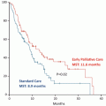
Then, they pointed out that population screening after implementing the LDCT screening in the public should confer similar benefits over time and minimizes risk as shown in the NLST study. Although it was solely a retrospective study, Nawa et al. disclosed a 24 % reduction (95 % CI of 14–33 %) in lung cancer mortality in a small district in Japan where lung cancer screening with LDCT had been implemented as a local government policy for 8 years [44]. Providing such data after implementing LDCT screening in the USA would further validate the usefulness of the screening.
- 1.
Eligible individuals must be accurately identified for screening as to age, smoking history, and willingness to adhere to a long-term screening program and undergo additional diagnostic procedures and treatment when necessary.
- 2.
The screening must be performed as part of a cohesive screening program to enhance its likelihood of success, and the cohesive program includes an adherence to evidence-based careening with technical parameters for LDCT, criteria for radiologists and imaging center, the use of a standardized nodule-identification-and-reporting system, smoking cessation program, follow-up evaluation, and central registration.
- 3.
Multidisciplinary involvement during the entire course from screening to treatment [43].
2.4 Future Directions Beyond CT Screening
2.4.1 Computer-Aided Evaluation of CT Screening
One of the shortcomings of the NLST was a high positive rate and high false-positive rate throughout the screening. As they classified all noncalcified pulmonary nodules ≥4 mm as positive, the positive rate was as high as 24.2 % [45]. It would bring a substantial harm for individuals who were screened if the screening with a positive rate of 24.2 % were implemented as a health-care policy. Although the final results from the NELSON study have not been disclosed, results of a prespecified analysis assessing screening performance in a subset population (n = 7155) were published [46]. In this screening process where semiautomated volumetric evaluation of pulmonary nodules with software was utilized, screening results were initially classified as negative, indeterminate, or positive based on nodule presence and volume. Subsequently, individuals with an initial indeterminate result underwent follow-up screening to classify their final screening result as negative or positive based on volume doubling time calculated by the software. That is to say, noncalcified pulmonary nodules ≥500 mm3 in volume, nodule volume doubling time <400 days plus increase in volume by ≥25 %, and newly appeared solid part in nonsolid nodules were judged as positive. Consequently, in contrast to the high positive rate (24.2 %) in the NLST, the positive rate was 2–3 %, and other parameters were excellent enough with sensitivity of 84.6 % (95% CI. 79.6–89.2), specificity of 98.6 % (95% CI, 98.5–98.8), positive predictive value of 40.4 % (95% CI, 35.9–44.7), and negative predictive value of 99.8 % (95% CI, 99.8–99.9). Although careful attention is needed because these data are still premature in contrast to the results from the NLST, comparison of the results between them seemed valuable as shown in Table 2.6. Computer-aided diagnosis (CAD) specific for lung cancer screening with LDCT has been developed to facilitate high sensitivity of LDCT screening without enforcing human labor burden [47, 48]. Although not fully established, a CAD equipped with automated volumetric software may further enhance sensitivity and suppress false-positive rate, resulting in excellent true positive predictive value.
Table 2.6
 Small Cell Lung Cancer and Molecular Targeted Therapy
Management of Adverse Effects by Molecular Targeted Therapy and Immunotherapy
Small Cell Lung Cancer and Molecular Targeted Therapy
Management of Adverse Effects by Molecular Targeted Therapy and Immunotherapy
 Minor-Driver Mutant
 Health-Related Quality of Life in Molecular Targeted Therapy
 EGFR Mutant
 PET-CT, Bio-imaging for Predicting Prognosis and Response to Chemotherapy in Patients with Lung Cancer


Comparison of major outcome parameters between the NLST and a subset of the NELSON
Related posts:
Small Cell Lung Cancer and Molecular Targeted Therapy
Management of Adverse Effects by Molecular Targeted Therapy and Immunotherapy
Minor-Driver Mutant
Health-Related Quality of Life in Molecular Targeted Therapy
EGFR Mutant
PET-CT, Bio-imaging for Predicting Prognosis and Response to Chemotherapy in Patients with Lung Cancer
Stay updated, free articles. Join our Telegram channel
Full access? Get Clinical Tree
