Sarcomas and Soft Tissue Tumors of the Head and Neck


Sarcomas and Soft Tissue Tumors of the Head and Neck


Marcus M. Monroe

Beverly A. Guadagnolo

Vinod Ravi

Wei-Lien Wang

Erich M. Sturgis


Sarcomas of the head and neck are a varied group of malignancies with widely differing clinical courses and clinical outcomes. Head and neck sarcomas arise from mesenchymal elements within the head and neck, including deep soft tissues, bone, and cartilage. Approximately 15% of all sarcomas arise within the head and neck, yet only 1% of all head and neck malignancies are sarcomas.1,2

Sarcomas are frequently categorized by their cell of origin and are generally broadly grouped into soft tissue sarcomas and bone/cartilaginous. More than 50 subtypes of sarcomas defined on the basis of histologic are recognized by the World Health Organization (WHO) (Table 27.1).3 Alternatively, sarcomas may be categorized according to their histologic grade, which may inform treatment decisions more directly than cell of origin does. For example, high-grade sarcomas are more likely to require complex multidisciplinary treatment plans.

For the vast majority of sarcomas, the etiology of the tumor is not known. Some genetic and environmental factors have been associated with development of sarcomas. For example, although very rare, several inherited syndromes are well known to increase the risk of specific types of sarcomas (Table 27.2). Germline mutations in the tumor suppressor gene TP53, such as those occurring in Li-Fraumeni syndrome, have been linked to an increased risk of both soft tissue and osteosarcomas.4,5 Diamond-Blackfan syndrome, a hereditary anemia linked to mutations in genes encoding ribosomal proteins, has been associated with osteosarcoma.6,7 Malignant fibrous histiocytoma (MFH) arising in bone occurs in up to 35% of patients with the rare cancer syndrome, diaphyseal medullary stenosis with MFH, and is characterized by loss of heterozygosity at the chromosomal band 9p21-22.8 Werner syndrome, characterized by rapid aging and mutations in the WMN gene, is well known to be associated with an increased incidence of multiple sarcoma types.9 Gardner syndrome, characterized by APC mutations and hereditary colon polyposis and cancer, is associated with an increased incidence of desmoid fibromatoses.10

The best characterized environmental risk factors linked to sarcoma development include the link between HIV infection and HHV8-induced Kaposi sarcoma (KS). Rarely, sarcoma can arise in the setting of prior exposure to ionizing radiation. Less well-established associations have been documented between increased sarcoma risk and environmental exposure to chlorophenols, herbicides, and certain chemicals in industrial waste products.11


PRINCIPLES OF SARCOMA MANAGEMENT


Patient Evaluation

Symptoms and clinical presentations in patients with sarcoma vary widely according to the histologic subtype and tumor location, but several sarcoma types have characteristic presentations. For instance, angiosarcomas typically present as a violaceous plaque on the scalps of elderly white males, whereas a chondrosarcoma often presents as a destructive bony lesion centered on the cricoid. The majority of types of sarcoma, however, lack a specific characteristic presentation and frequently present with an asymptomatic mass.11 In a review of over 2,000 patients presenting to MD Anderson Cancer Center over the past 44 years (Tables 27.3 and 27.4), the most frequent locations for sarcomas arising in the head and neck of adults (Table 27.3) included the scalp/face (30% of cases), followed by the sinonasal cavity/anterior skull base (23%) and the upper aerodigestive tract and the parotid/neck (each accounting for 19%). Sarcomas arising in the ear/lateral or posterior skull base were quite rare (<8%). In the pediatric and adolescent population (Table 27.4), the distribution of sites is somewhat different with the sinonasal cavity/anterior skull base accounting for 28% of cases and the upper aerodigestive tract for 27% followed by the scalp/face (22%), the parotid/neck (14%), and the ear/lateral or posterior skull base (9%).

The physical examination in a patient with a known or suspected sarcoma should entail a careful assessment of the extent of the disease, the degree of fixation to surrounding structures, and the presence or absence of regional adenopathy. Cross-sectional imaging should be performed to further delineate the extent of the primary lesion and to assess for regional and distant spread of disease. For most head and neck sarcomas, distant metastases are most likely to present in the lungs, and chest imaging with computed tomography should be obtained routinely. For sarcomas with a higher risk of intracranial metastases, such as alveolar soft part sarcoma (ASPS) and angiosarcoma, the staging workup should include imaging of the brain.12

Although fine needle biopsy is the initial biopsy of choice for many head and neck masses, biopsy for head and neck sarcomas often entails either a core needle or incisional biopsy as the limited amount of tissue and lack of tissue architecture provided by fine needle aspiration makes diagnosis of
sarcomas particularly difficult. Biopsy sites should be planned such that they can be easily excised during subsequent surgical resection. Pathologic assessment of the biopsy specimen should be undertaken by a pathologist with experience in sarcomas13 with the goal of providing information on the histologic subtype as well as tumor grade at a minimum. Standard morphologic assessment remains the standard for diagnosis, although the use of cytogenetics to augment standard morphologic assessment is accelerating (Table 27.1). For consistency and communication among providers, pathologic reports should use the nomenclature for sarcomas standardized by the WHO.3 Approximately one-quarter of head and neck sarcomas arising in adults are unclassified and undifferentiated, high-grade, pleomorphic sarcomas, with osteosarcomas, angiosarcomas, and rhabdomyosarcomas accounting for another one-third (Table 27.3). However, in children and adolescents, the majority are rhabdomyosarcomas (Table 27.4).


Table 27.1 Etiologic Factors for Head and Neck Sarcomas


Genetic Predisposition


Gene(s)


Sarcoma(s)


Li-Fraumeni syndrome


TP53


Bony and soft tissue sarcomas


Hereditary retinoblastoma


Rb


Osteosarcoma


DMS-MFH


LOH at 9p21-22


Malignant fibrous histiocytoma (MFH)


Werner syndrome


WMN


Bony and soft tissue sarcomas


Gardner syndrome


APC


Desmoid tumors


Neurofibromatosis type 1


NF1


MPNST


Costello syndrome


HRAS


Rhabdomyosarcoma


Beckwith-Wiedemann syndrome


CDKN1, IGF2, H19, KCNQ1OT1


Rhabdomyosarcoma


Rothmund-Thompson syndrome


RECQL4


Osteosarcoma


Paget disease


Unknown


Osteosarcoma


Ollier and Maffucci syndromes


IDH1, IDH2


Chondrosarcoma


Environmental Exposures


Sarcoma(s)


Ionizing radiation


Bony and soft tissue sarcomas


HIV, HHV-8


Kaposi Sarcoma


Industrial waste


Bony and soft tissue sarcomas


Herbicides and chlorophenols


Bony and soft tissue sarcomas


DMS-MFH, diaphyseal medullary stenosis with malignant fibrous histiocytoma; HLRCC, hereditary leiomyomatosis and renal cell cancer; MPNST, malignant peripheral nerve sheath tumors.


Staging and Prognostic Factors in Head and Neck Sarcomas

The American Joint Committee on Cancer staging system for soft tissue sarcomas incorporates measures of tumor size (> or ≤5 cm), location with respect to deep fascial planes, the presence of regional or distant metastatic spread, and tumor grade. For sarcomas located in the head and neck, the usefulness of this staging system has been questioned, as such tumors are typically smaller than 5 cm at the time of presentation and have high rates of deep fascial involvement.1,14

For most histologic subtypes of sarcoma, studies have shown that location within the head and neck portends a worse prognosis than location in other anatomic regions.15 Tumor grade and margin status remain the most consistently identified prognostic factors in multiple case series of head and neck sarcomas.1,16,17,18 Unfortunately, the proximity to vital structures frequently precludes wide resection margins for head and neck sarcomas, a factor that has been linked to their poor prognosis. In addition to grade and margin status, tumor size, age, and smoking status have been associated with reduced survival.15,16,17,18,19 The prognostic significance of regional metastatic spread for head and neck sarcomas is debated.14,20


Principles of Surgery

The traditional cornerstone of therapy for head and neck soft tissue and bony sarcomas without evidence of distant metastatic disease on presentation is surgical excision with histologically negative margins. Given the propensity for microscopic spread beyond the clinically apparent tumor, margins of 2 cm and inclusion of an adjacent normal-appearing tissue plane beyond the mass should be attempted. Surgical resections should be planned to include excision of the biopsy site. Within the head and neck, proximity of critical structures frequently precludes obtaining wide margins. If close or histologically positive surgical margins are anticipated on the basis of preoperative clinical and radiologic evaluation, neoadjuvant chemotherapy and/or preoperative radiation therapy should be considered.12 Standard terms used to describe surgical margins for sarcomas are R0 for microscopically negative margins, R1 for microscopically positive margins, and R2 for grossly positive margins.

Head and neck sarcomas should be managed by experienced multidisciplinary teams with experience in treating sarcomas. Surgical management of head and neck sarcomas frequently involves multispecialty surgical teams including thoracic, vascular, reconstructive, ophthalmologic, and neurosurgical services, but regardless of which other specialists are involved, an experienced head and neck surgeon is essential.
Given the frequent need for adjuvant chemotherapy and radiation therapy either before or after surgery, close coordination with medical and radiation oncologists with experience treating sarcomas is critical.


Table 27.2 Histologic Subtypes of Head and Neck Sarcomas with Known Genetic Alterations


WHO Sarcoma Histologic Subtypes


Cytogenic Alterations


Altered Gene(s)


Adipocytic Tumors


Dedifferentiated liposarcoma


Supernumerary ring and giant marker chromosomes, amplification 12q13-15


MDM2, CDK4, HMGA2, SAS, GL1


Myxoid/round cell liposarcoma


t(12;16)(q13;p11), t(12;22)(q13;q12)


FUS-DD1T3, EWSR1-DD1T3


Pleomorphic liposarcoma


Complex alterations


Fibroblastic, Myofibroblastic, and Fibrohistiocytic Tumors


HPC/SFTs


Complex alterations


NAB2-STAT6


Fibrosarcoma


Complex alterations


Myxofibrosarcoma


Complex alterations


Low-grade fibromyxoid sarcoma


t(7;16)(q33;p11), t(11;16)(p11;p11)


FUS-CREB3L2, FUS-CREB3L1


Sclerosing epithelioid fibrosarcoma


EWSR1-CREB3L1


Undifferentiated pleomorphic sarcoma/MFH


Desmoid fibromatosis


Trisomy 8 or 20; Loss of 5q21


CTNNB1 or APC mutations


DFSPs


t(17;22)(q21;q13) and derivative ring chromosomes


COL1A1-PDGFB


Smooth Muscle Tumors


Leiomyosarcoma


Complex alterations


Skeletal Muscle Tumors


Embryonal rhabdomyosarcoma


LOH at 11p15


Alveolar rhabdomyosarcoma


t(2;13)(q35;q14), t(1;13)(p36;q14), t(X;2) (q13;q35)


PAX3-FKHR and PAX7-FKHR


Pleomorphic rhabdomyosarcoma


Complex alterations


Vascular Tumors


Epithelioid hemangioendothelioma


t(1;3)(p36.3;q25)


Angiosarcoma


Complex alterations


PTPRB and PLCG1 mutations


Tumors of Peripheral Nerves


Malignant peripheral nerve sheath tumors


Complex alterations


Tumors of Uncertain Differentiation


Synovial sarcoma


t(X;18)(p11;q11)


SS18-SSX1, SS18-SSX2, SS18-SSX4


Alveolar soft-part sarcoma


der(17)t(X;17)(p11;q25)


ASPL-TFE3


Sarcomas of Cartilage and Bone


Chondrosarcoma


Complex alterations


Osteosarcoma


Complex alterations


PNET/Ewing sarcoma


t(11;22)(q24;q12), t(21;22)(q22;q12), t(2;22) (q33;q12), t(7;22)(p22;q12), inv(22)(q12;q12), t(16;21)(p11;q22)


EWS-ETS


Principles of Radiation Therapy

For head and neck sarcomas, preoperative or postoperative adjuvant radiation therapy is typically given as part of combined modality primary local therapy for nonmetastatic presentations. Multiple case series have demonstrated improved local control with the use of adjuvant radiation therapy in cases of positive margins or high-grade tumors.21,22 Unfortunately, negative margins can be difficult to achieve in head and neck sarcoma surgery; in some series, up to half of patients have R1 or R2 resections.11 Low-grade sarcomas with wide negative margins may be considered for single-modality treatment with surgery alone.

Although the benefits of adjuvant radiation therapy for sarcomas in the head and neck are widely accepted, the optimal timing of radiation therapy remains controversial. Both preoperative and postoperative radiation therapies have potential advantages. Tumor margins and the regions that need to be targeted with radiation are more easily defined before surgical resection, when the sarcoma mass is present during simulation.
Consequently, preoperative radiation therapy has the advantages of a lower dose (50 Gy vs. 60 to 66 Gy for postoperative radiation therapy) and a smaller, precisely defined radiation field, translating into a lower risk of late radiation-related side effects, including fibrosis and edema, and the potential to minimize the radiation dose to critical structures.23 However, preoperative radiation therapy has been reported to be associated with increased wound complications following surgery. In a randomized trial that attempted to address this issue, 190 patients with soft tissue sarcoma of the extremities were randomized to preoperative or postoperative adjuvant radiation therapy.24 Similar rates of local control, disease-free survival, disease-specific survival, and overall survival were noted between the groups. Among patients with lower extremity sarcomas, wound complications were more common in patients undergoing preoperative radiation therapy. While late side effects of radiation therapy are presumed to be lower with lower doses used preoperatively then with postoperative doses, long-term side effects have not been reported for this trial. In a separate analysis of 40 consecutive patients with head and neck soft tissue sarcomas treated with preoperative radiation therapy, the rate of wound complications was lower than that reported previously for soft tissue sarcomas of the extremities,25 suggesting that the improved blood supply of the head and neck may reduce the risk of some of the wound complications associated with preoperative radiation therapy at other sites. The use of preoperative radiation therapy should be considered in all cases of sarcoma of the head and neck where radiation will be used and the complication risk for surgery in an irradiated field does not preclude it.26 This requires treatment plan coordination and discussion between the treating radiation oncologist and the head and neck surgeon.


Table 27.3 Adult Head and Neck Sarcomas at the University of Texas MD Anderson Cancer Center (Age ≥ 18, 1970-2013)


Histologic Type


No.


%


Site of Origin


Scalp and Face


Sinonasal Tract Anterior/Medial Skull Base


Ear Lateral/Posterior Skull Base


Upper Aerodigestive Tract


Parotid and Neck


Bone and Cartilage


Osteosarcoma


288


14.8


3


108


48


125


4


Ewing sarcoma/PNET


35


1.8


5


10


9


5


6


Chondrosarcoma


153


7.9


2


45


52


46


8


Fibrous


Undifferentiated pleomorphic sarcoma/MFH


159


8.2


59


27


10


17


46


Fibrosarcoma


49


2.5


10


16


0


11


12


Fibromyxoid sarcoma


4


0.2


1


0


0


0


3


DFSP


110


5.7


89


0


1


0


20


Muscular


Rhabdomyosarcoma


199


10.3


22


112


1


52


12


Leiomyosarcoma


72


3.7


17


14


2


15


24


Vascular


Angiosarcoma


240


12.4


209


8


4


6


13


Hemangiopericytoma


53


2.7


11


21


2


7


12


Kaposi sarcoma


37


1.9


17


0


1


6


13


Neural


Neurogenic sarcoma


72


3.7


18


7


0


4


43


Fatty


Liposarcoma


38


2.0


7


4


0


5


22


Histogenesis Unclear


Synovial sarcoma


72


3.7


14


3


1


21


33


Alveolar soft part sarcoma


12


0.6


2


3


0


4


3


Unclassified and Other


348


17.9


99


70


21


55


103


Head and Neck Sarcoma (Totals)


1941


100


585


448


152


379


377


Principles of Chemotherapy

For sarcomas that display evidence of distant metastatic spread, chemotherapy is generally indicated and is individualized on the basis of specific tumor histologic features.26 For localized resectable sarcomas in adults, the role of chemotherapy remains
less well defined. The Sarcoma Meta-analysis Collaboration analyzed results from 14 trials evaluating doxorubicin-based therapy in resectable soft tissue sarcomas. Local, distant, and overall recurrence-free survival times were significantly improved with doxorubicin-based adjuvant chemotherapy, although the absolute improvement in overall recurrence-free survival was only 6% to 10% at 10 years.27 Other trials have demonstrated histologic subtype-specific responses to different chemotherapeutic agents and are discussed below. Authors increasingly describe the use of chemotherapy to treat sarcomas, particularly the use of neoadjuvant chemotherapy for high-risk tumors.


Table 27.4 Pediatric Head and Neck Sarcomas at the University of Texas MD Anderson Cancer Center (Age < 18, 1970-2013)


Histologic Type


No.


%


Site of Origin


Scalp and Face


Sinonasal Tract Anterior/Medial Skull Base


Ear Lateral/Posterior Skull Base


Upper Aerodigestive Tract


Parotid and Neck


Bone and Cartilage


Osteosarcoma


30


7.5


0


1


8


21


0


Ewing sarcoma/PNET


25


6.3


5


5


8


4


3


Chondrosarcoma


10


2.5


0


6


3


1


0


Fibrous


Undifferentiated pleomorphic sarcoma/MFH


8


2.0


3


1


2


1


1


Fibrosarcoma


10


2.5


5


0


0


3


2


Fibromyxoid sarcoma


1


0.3


0


0


0


1


0


DFSP


5


1.3


5


0


0


0


0


Muscular


Rhabdomyosarcoma


228


57.6


60


82


13


54


19


Leiomyosarcoma


2


0.5


0


1


0


1


0


Vascular


Angiosarcoma


1


0.3


0


0


0


1


0


HPC


4


1.0


1


1


1


0


1


Neural


Neurogenic sarcoma


13


3.3


4


0


1


0


8


Fatty


Liposarcoma


2


0.5


0


1


0


1


0


Histogenesis Unclear


Synovial sarcoma


9


2.3


0


0


0


3


6


Alveolar soft part sarcoma


6


1.5


0


1


0


3


2


Unclassified and other


42


10.6


6


12


1


11


12


Head and neck sarcoma (totals)


396


100


89


111


37


105


54


SOFT TISSUE SARCOMAS


Soft Tissue Tumors of Endothelial Origin


Angiosarcoma

Angiosarcomas are rare soft tissue sarcomas arising from endothelial cells. They classically present as a violaceous macule or papule on the scalp in elderly white males, although they can present in people of any age, gender, or ethnicity and in any anatomic location. Most angiosarcomas in the head and neck arise spontaneously in the elderly, although several well-known risk factors are associated with the development of angiosarcomas at other sites in the body. These risk factors include chronic lymphedema following treatment for breast cancer (Stewart-Treves syndrome), prior radiation exposure, and prior exposure to environmental toxins such as vinyl chloride, thorium dioxide, anabolic steroids, arsenic, and radium.28 Susceptibility to angiosarcoma has been proposed to be increased in people with mutations in BRCA1 and BRCA2, neurofibromatosis, Maffucci syndrome, and Klippel-Trenaunay syndrome. Up to 38% of cases of angiosarcoma harbor mutations in PTPRB or PLCG1.29

Frequent delays in diagnosis are common for head and neck angiosarcomas given their propensity to mimic benign conditions, including bruises and benign vascular lesions, typically of the scalp (Fig. 27.1, Table 27.3). Angiosarcomas, particularly those of the scalp, have a propensity for diffuse spread beyond the visible cutaneous abnormality, making underestimation of the true extent of disease commonplace. Satellite lesions occur
in up to half of patients and portend a worse prognosis.30 Regional nodal disease at presentation is relatively uncommon, occurring in 6% to 10% of patients,31 although up to 16% of patients may experience delayed regional recurrence following treatment.30 The development of distant metastatic disease occurs in 36% to 77% of patients,30,31,32 and the most common site of metastasis is the lungs. Local relapse occurs in 35% to 53% of patients30,31,32 and is more common with larger tumor size, tumor location on the scalp, and single-modality treatment.30,33,34 Local relapse portends a poor prognosis.30


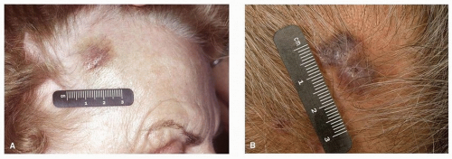
Figure 27.1. Angiosarcoma of the right temple (A) and scalp (B). Early lesions may mimic that of benign conditions such as bruising, leading to delays in diagnosis.

Angiosarcomas arising in the head and neck have a worse prognosis than do those arising in other sites: the estimated 5-year survival rate for scalp and neck angiosarcomas is 34%, compared to 64% to 75% for those arising at other locations.35 Among angiosarcomas of the head and neck, angiosarcoma of the scalp is associated with worse disease-specific survival than are tumors at other locations.30

The combination of surgery and radiation therapy appears to offer the best chance for local control as well as improved disease-specific and overall survival as compared to either surgery alone or radiation alone.30,31,33,34 Achieving an R0 resection can be particularly difficult in angiosarcomas of the head and neck given the propensity for a diffuse and multifocal growth pattern.30,33 Because of this, some have argued against extensive resections necessitating complicated reconstructions that may delay postoperative radiation therapy.30 Given the need for adjuvant radiation therapy in nearly all cases of angiosarcoma, repair of surgical defects should be robust enough to withstand radiation therapy. In particular, skin grafting for repair of scalp defects should be avoided when possible. Limited evidence exists to support the role of chemotherapy for localized angiosarcomas, although neoadjuvant taxane-based chemotherapy followed by radiation therapy and/or limited surgical excision has produced encouraging results.30,36,37

Only gold members can continue reading. Log In or Register to continue

Stay updated, free articles. Join our Telegram channel

Dec 18, 2016 | Posted by in ONCOLOGY | Comments Off on Sarcomas and Soft Tissue Tumors of the Head and Neck

Full access? Get Clinical Tree

Get Clinical Tree app for offline access