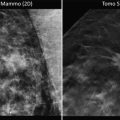
Fig. 7.1
(a) First stereotactic table imported into the United States in 1985. (b) The module on which the biopsy needle was positioned and manually operated. (c) Stereotactic breast biopsy needles used for sampling breast lesions. The two used at Karolinska and the University of Kiel, West Germany, retrieved cytologic samples. The 18-gauge needle was at that time used for renal biopsies and was adapted for breast lesions
The accuracy of the test and the follow-up results were satisfactory provided an experienced cytopathologist was available, but was suboptimal otherwise [7]. The author then began taking core samples with a 20-gauged needle in a consecutive series of 250 patients who were scheduled to undergo open biopsy for suspicious mammograms. The results showed sensitivity of 71 %, specificity of 96 %, and insufficiency rate of 17 % [8]. Other investigators employed 14-gauged needles with improved results [9–11].
“Needle instead of knife” approach for diagnosis of mammographic abnormalities was presented to several local and national surgical meetings but it was not received with enthusiasm. Radiologists, on the other hand, accepted the technology immediately and began exploring its application on a large scale and dominating the field. A decade later general/breast surgeons appealed to the American College of Surgeons to organize training courses for stereotactic needle biopsy training, which started in 1998 in Chicago and presently continues at different sites by the American College of Breast Surgeons. Currently radiologists perform the majority of stereotactic needle biopsies nationally and consider the procedure as part of their specialty. A formal action was initiated through the Institute of Medicine, Congress, and FDA to block surgeons from performing this procedure. At the community level the executive health care committees hesitate to grant trained surgeons the priviledge of practicing the procedure. The issue is an ongoing dispute at this time.
Modern Stereotaxis
Since its inception in Sweden and introduction into the United States, stereotactic technology has undergone several major changes. The first one was the introduction of digital imaging [12, 13]. This change was incorporated in the stereotactic devices even before their application in routine mammography. The change facilitated the biopsy procedure significantly by shortening the operation time. Furthermore, the high resolution capability together with magnification and rapid image processing features enabled the operator to visualize minute parenchymal changes for sampling. The second development was computerization of the system which allowed rapid transfer of the data from the monitor screen to the mechanized platform. Third was the emergence of vacuum-assisted cutting probes which once appropriately positioned in the breast removed multiple samples from the targeted lesion with one instead of multiple passes as was the case with mechanized needles [14, 15]. Thus with the combination of high-resolution digital screening mammography resulting in detection of smaller cancers and larger needles removing 10–15 core samples, one is witnessing complete percutaneous removal of some breast tumors as documented on ensuing excisional biopsy.
Other Breast Stereotactic Technologies
In late 1980s Fischer Corporation based in Denver CO acquired the license and production rights of the stereotactic table in the United States. Soon afterwards LoRad/Hologic Corp. in Danbury CT introduced their version of breast stereotactic table whereby the biopsy unit including the X-ray arm could be rotated 180° around the breast, thus enabling the operator to biopsy the lesions from all directions. This was a technical advantage partially cancelled by slight disadvantage of the technology being based on Cartesian principle, meaning that the platform could be raised parallel with the table undersurface, limiting visualization of lesions in the far posterior 2–5 mm part of the breast close to the chest wall because the X-ray beam travels parallel with the undersurface of the table. This problem could in some cases be resolved, in rare occasions, by bringing the patient’s arm through the table opening.
Both Fischer and LoRad tables have to be secured onto the floor although their “mobile” versions can be transported to a medical facility in an especially equipped truck on a prearranged date for performance of breast biopsies. In 2006, Siemens introduced yet another stereotactic table into clinical practice. This was an upright device which could be attached to a regular mammographic unit, thus lowering the cost and obviating the need for a dedicated space. The biopsy is done with the patient sitting upright, which is an advantage as lying prone on the traditional table for up to an hour causes neck discomfort in some patients. On the other hand the dedicated table allows the gravity to pull the breast away from the chest wall allowing access to the posterior targets. Additionally, patients are unlikely to faint in a prone position and the operating surgeon has a better sense of control.
Role of Ultrasound in Diagnosis of Breast Lesions
During the past decade there has been a dramatic improvement in the resolution of breast sonography. Soft tissue densities as small as 5 mm in diameter and even clusters of microcalcifications may be visualized by ultrasound. Miniaturization of the technology has made the device small enough to be portable and to be used in the office, at the bedside, as well as in the operating room. Ultrasound differentiates cystic from solid breast lesions. Its acoustic properties demonstrate homogeneity of the lesion, thus differentiating benign from malignant tumors. Additionally, color Doppler ultrasound demonstrates the vascularity of the suspected tumor.
Ultrasound has truly become the 21st-century stethoscope of the surgeon. In clinical practice ultrasound-guided needle biopsy has replaced most breast biopsies previously performed on a stereotactic table [16]. The emergence of three-dimensional ultrasound makes this breast imaging technology even more appealing.
Role of “Functional Imaging” in Diagnosis of Breast Lesions
1.


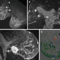
Magnetic resonant imaging (MRI) is a technology which has come to play a significant role in breast imaging especially in dense breasts of younger women where mammography is not sensitive enough to detect small tumors at early phases of development [17–19]. In today’s practice, MRI is deployed in patients with dense breasts to rule out multi-centricity as well as bilaterality once a focus of malignancy is documented in one breast. MR-guided breast biopsy is increasingly practiced in younger women with dense parenchyma and in patients with scars of previous interventions.
Stay updated, free articles. Join our Telegram channel

Full access? Get Clinical Tree


