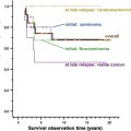
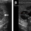
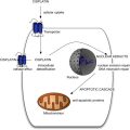
Advanced germ cell tumors (GCTs) are curable with the appropriate integration of cisplatin-based chemotherapy and postchemotherapy surgical resection of residual masses. For men with retroperitoneal metastases, postchemotherapy retroperitoneal lymph node dissection (PC-RPLND) is a vital component of this treatment algorithm. The rationale for PC-RPLND is based on the consistent 10% to 20% and 35% to 55% incidence of viable malignancy and teratoma, respectively. Prognostic factors and nomograms cannot predict the presence of necrosis with sufficient accuracy to obviate the need for PC-RPLND. This article reviews the indications, technique, and outcomes of PC-RPLND in the management of advanced GCT.
Testis cancer is the most common malignancy afflicting men between the ages of 15 and 34 years, with 8400 men expected to be diagnosed in the United States in 2010. Most cases (>95%) are germ cell tumors (GCTs), which are broadly divided into seminoma and nonseminoma GCT (NSGCT). Advanced GCT is an example of the importance of multidisciplinary management in the successful treatment of patients. Before the development of cisplatin-based chemotherapy, long-term survival was reported in less than 10% of patients. Long-term cure is now anticipated in 80% to 90%. A recent meta-analysis of 10 trials enrolling a total of 1775 patients with advanced NSGCT reported improved 5-year survival rates for patients with good-risk (94% vs 89%), intermediate-risk (83% vs 75%), and poor-risk disease (71% vs 41%) compared with the original pooled analysis conducted by the International Germ Cell Cancer Collaborative Group (IGCCCG). This improved prognosis for patients with advanced GCT is likely caused by better risk stratification and risk-appropriate chemotherapy, stage migration, and improved integration of chemotherapy and postchemotherapy surgery (PCS) for resection of residual masses.
For treatment purposes, the distinction between seminoma and NSGCT holds great importance, particularly in the management of residual masses. Compared with NSGCT, seminoma is exquisitely sensitive to cisplatin-based chemotherapy. Thus, residual masses after first-line chemotherapy for seminoma are more likely to show necrosis and less likely to harbor viable GCT elements compared with NSGCT. The risk of teratoma at metastatic sites has a substantial effect on treatment algorithms for NSGCT and necessitates the frequent use of PCS in patients with advanced NSGCT. As discussed later, teratoma is not sensitive to chemotherapy and the outcome of patients with metastatic teratoma is related to the completeness of surgical resection. Although histologically benign, teratoma has unpredictable biology, with a capacity to grow rapidly, undergo malignant transformation, or result in late relapse. The risk of teratoma at metastatic sites is generally not a consideration for advanced seminoma, which has important implications for the management of residual masses after chemotherapy.
Because of the high cure rates anticipated for patients with advanced GCT, numerous clinical trials have been conducted in an attempt to minimize treatment and avoid any unnecessary therapies in an effort to reduce short-term, and particularly long-term, toxicity. One such approach has been to limit the number of patients who receive 2 interventions (double therapy): either surgery or chemotherapy and not both. However, because NSGCTs are usually mixed tumors and teratoma often exists at metastatic sites with other GCT elements, cure often requires chemotherapy to eradicate the chemosensitive components and surgery to remove teratomatous components. It is widely accepted that the successful integration of systemic therapy and PCS is a major contributing factor to the improved cure rates for metastatic GCT seen in the past several decades.
Advances in surgical technique and understanding of retroperitoneal anatomy have reduced the morbidity of RPLND (eg, retrograde ejaculation) while enhancing oncological efficacy. However, postchemotherapy retroperitoneal lymph node dissection (PC-RPLND) can be a challenging undertaking that has historically been associated with higher rates of complications compared with primary RPLND. As such, appropriate patient selection and proper surgical technique are critical to optimizing cancer and quality-of-life outcomes following surgery.
The indications and outcomes of PC-RPLND are discussed to determine its role in the contemporary management of advanced GCT. This article focuses primarily on PC-RPLND in the setting of NSGCT, but the limited indications for PCS in advanced seminoma are also discussed.
NSGCT: rationale for PC-RPLND
The role of PC-RPLND for residual masses in advanced NSGCT is well established and its rationale is based on several factors. Multiple large series of patients undergoing PC-RPLND for residual masses after first-line chemotherapy have consistently reported evidence of persistent GCT elements in the resected specimens in 50% or more of patients. On average, histopathologic evaluation of resected specimens shows necrosis, teratoma, and viable malignancy (with or without teratoma) in 40%, 45%, and 15% of cases respectively ( Table 1 ).
| Patients | Necrosis (%) | Viable Malignancy ± Teratoma (%) | Teratoma Only (%) | |
|---|---|---|---|---|
| Steyerberg et al, 1995 | 556 | 45 | 13 | 42 |
| Carver et a, 2007 | 504 | 49 | 11 | 39 |
| Hendry et al, 2002 | 330 | 25 | 9 | 66 |
| Debono et al, 1997 | 295 | 25 | 7 | 67 |
| Spiess et al, 2006 | 236 | 41 | 17 | 42 |
| Albers et al, 2004 | 232 | 35 | 31 | 34 |
| Toner et al, 1990 | 185 | 47 | 16 | 37 |
| Steyerberg et al, 1998 | 172 | 45 | 13 | 42 |
| Stenning et al, 1998 | 153 | 29 | 15 | 55 |
| de Wit et al, 1997 | 127 | 35 | 9 | 56 |
| Oeschle et al, 2008 | 121 | 45 | 21 | 34 |
| Sonneveld et al, 1998 | 113 | 46 | 9 | 45 |
| Gerl et al, 1995 | 111 | 47 | 12 | 41 |
| Hartmann et al, 1997 | 109 | 52 | 21 | 27 |
The therapeutic benefit of PC-RPLND in cases in which residual masses harbor viable malignancy or teratoma is well documented. Complete resection of viable malignancy (with or without adjuvant chemotherapy) is associated with 5-year survival rates between 45% and 77%. If not excised, or subject to an inadequate resection, residual viable malignancy within the retroperitoneum will presumably relapse, necessitating second-line (salvage) chemotherapy; however, only a quarter of such relapsing patients experience long-term survival.
Although histologically benign, the risk of teratoma within residual masses also mandates complete surgical excision, which is associated with long-term cancer control in 75% to 90% of men. Teratoma is resistant to chemotherapy and radiation therapy and the natural history of unresected teratoma after chemotherapy is unpredictable. It may remain dormant or exhibit slow growth. However, it may also exhibit rapid, expansive, local growth, leading to invasion or impingement of adjacent vital structures. Moreover, a small percentage (typically <10%) of teratomas may undergo malignant transformation to a non-GCT malignancy (eg, rhabdomyosarcoma, primitive neuroectodermal tumor, adenocarcinoma), which is resistant to conventional GCT chemotherapy regimens and is associated with a poor prognosis. Lastly, unresected teratoma may manifest as late recurrence, which is associated with poor prognosis. As with viable malignancy, complete eradication of all residual teratomatous elements following chemotherapy is the best means of optimizing oncological outcomes.
The critical role of PC-RPLND in advanced GCT is shown by the results of a recent randomized trial comparing BEPx3 versus EPx4 in 257 men with good-risk, advanced NSGCT. In this trial, the indications for, and technique of, PC-RPLND was not dictated by protocol, and fewer than 50% of study participants underwent PCS for residual masses. Overall, 14 of 20 relapses (70%) and 7 of 14 deaths (50%) occurred in patients who either did not undergo PCS or who relapsed in the retroperitoneum after PC-RPLND (presumably from a limited-template dissection).
Hendry and colleagues analyzed the outcomes of PC-RPLND in 330 and 112 patients after first-line and second-line chemotherapy, respectively. After first-line chemotherapy, patients were more likely to undergo a complete resection, resected specimens were less likely to contain viable malignancy, and the risk of relapse (17% vs 38%) and death (11% vs 44%) was substantially less than after second-line chemotherapy. These results suggest that a potential opportunity to cure patients may be lost if PC-RPLND is not performed following first-line chemotherapy.
Debono and colleagues analyzed the outcomes of 295 patients with NSGCT treated with first-line chemotherapy at Indiana University between 1987 and 1994. Patients with residual masses after chemotherapy who underwent PC-RPLND had excellent relapse-free survival (86%–87%), which approached that of patients who achieved a complete response to chemotherapy alone who were observed without PC-RPLND (92%). However, the former group had a substantially improved relapse-free survival compared with those who were observed after a major (but not complete) response to chemotherapy (74%) and those who had an incomplete surgical resection of residual masses (40%). Taken together, these studies show the potential role of PC-RPLND in reducing the risk of relapse and mortality from GCT.
Besides a therapeutic benefit, resection of residual masses can also provide staging information that can be used to dictate further chemotherapy and/or surveillance protocols. Extent of resection and percentage of viable malignancy in the tissue specimen have been shown to be important prognostic factors in predicting survival. Moreover, the presence of viable malignancy in residual masses suggests the need for more chemotherapy, although its use in this setting is controversial. In a study by Fox and colleagues, 14 of 27 patients (70%) found to have viable malignancy after undergoing PC-RPLND and who were treated with adjuvant chemotherapy were free of recurrence, compared with 0 of 7 patients who were observed. However, a multi-institutional study comprising 238 patients with viable malignancy in PC-RPLND specimens showed that postoperative chemotherapy was associated with improved 5-year progression-free survival (69% vs 52%, P <.001) but not overall survival (74% vs 70%, P = .7). A confirmatory analysis of 61 patients from the same investigators reported similar findings, with a 15-point improvement in progression-free survival but little difference in overall survival, but the study was not powered for survival analysis. The impact of postoperative chemotherapy in this setting thus remains unclear.
NSGCT: patient selection for PC-RPLND
PC-RPLND is generally recommended for patients with NSGCT with significant residual masses (ie, >1 cm) and normal serum tumor markers, because viable malignancy or teratoma is found in residual masses in approximately 15% and 40% of cases, respectively. Given that approximately 45% of patients have necrosis, some have argued against the use of PC-RPLND in all patients based on the assumption that those with necrosis do not derive any therapeutic benefit. Although patients with necrosis in resected specimens have a risk of relapse of 10% or less, it is conceivable that such favorable outcomes may be achieved without surgery. However, numerous desperation RPLND series have reported postoperative normalization of serum tumor marker levels even among patients with just necrosis in resected specimens. A recent study from Indiana University also reported that 33% of men with necrosis had evidence of molecular changes associated with GCT within stromal cells. These studies suggest that some residual viable GCT elements may be missed on routine histopathologic analysis, indicating a potential therapeutic benefit to PC-RPLND even among patients with necrosis. However, this concept is difficult to prove in the absence of a randomized trial.
The ability to accurately predict tissue histology and identify those men with only necrosis potentially obviates the need for PC-RPLND in select patients. Several clinical factors have been identified as predictors of necrosis in residual masses, including the absence of teratoma in the primary tumor, the percentage reduction in retroperitoneal tumor burden after chemotherapy, and the size of the retroperitoneal mass before and after chemotherapy. Nomograms have been developed to predict the presence of necrosis based on these and other factors. Although these nomograms discriminate well between patients with residual GCT versus those with necrosis only, they are associated with a false-negative rate of 20%, and thus cannot be used reliably to dictate therapy. Furthermore, no imaging modality has proved reliable in the prediction of the histology of residual masses. In a prospective study of 121 patients with residual masses after first-line chemotherapy, the predictive accuracy of positron emission tomography (PET) (56%) for viable malignancy or teratoma was no better than computed tomography (CT) (55%) or postchemotherapy serum tumor markers (56%). Based on these findings, no imaging modality or multiparameter nomogram can be used safely to exclude a man from PC-RPLND who has a residual mass greater than or equal to 1 cm after first-line chemotherapy.
The management of patients with complete serologic and radiographic response is controversial, with some guidelines advocating close observation and others recommending PCS if prechemotherapy mass size is greater than 3 cm. Some institutions advocate PC-RPLND in all patients after a complete response to first-line chemotherapy, regardless of the residual mass size, based on the rationale that there are no means to reliably exclude the presence of residual GCT and the risks associated with PC-RPLND are low when performed by experienced surgeons. A 1-cm cutpoint is arbitrary and numerous studies have shown that, on average, patients with residual masses 20 mm or smaller have a 30% and 6% incidence of teratoma and viable malignancy, respectively ( Table 2 ). However, 2 studies have reported a low risk of relapse (4%–10%) and 97% to 100% cancer-specific survival in patients with residual masses less than 1 cm who were observed without PCS. The highly select nature of these patients is shown by most being good-risk by IGCCCG criteria, 60% to 76% not having teratoma in the primary tumor, and many having no visible masses after chemotherapy. The median follow-up in 1 of the studies was only 3.8 years, and likely underestimates the risk of relapse and death. In a median follow-up of 15 years in the Indiana University study, half of the relapses occurred in the retroperitoneum. Thus, when deciding on PC-RPLND for small residual masses, the morbidity of PC-RPLND must be balanced with the risks of observation, including chemorefractory relapse and the frequent use of CT imaging in the surveillance of the retroperitoneum. Radiation from CT imaging may be an important cause of secondary malignancies and the safety threshold is exceeded after approximately 7 CT scans; routine CT imaging is not necessary after a full, bilateral template PC-RPLND. Observation is a reasonable strategy for only 25% or less of men with advanced NSGCT. At our institution, observation is restricted to men with IGCCCG good-risk disease, no evidence of teratoma in the primary tumor, no evidence of any residual mass following chemotherapy, and who are anticipated to be compliant with follow-up imaging and testing.
| Patients | Size (mm) | Necrosis (%) | Viable Malignancy ± Teratoma (%) | Teratoma Only (%) | |
|---|---|---|---|---|---|
| Steyerberg et al, 1995 | 275 | ≤20 | 65 | 5 | 30 |
| Steyerberg et al, 1995 | 162 | ≤10 | 72 | 4 | 24 |
| Oldenburg et al, 2003 | 87 | ≤20 | 67 | 7 | 26 |
| Fossa et al, 1992 | 78 | <20 | 68 | 4 | 29 |
| Fossa et al, 1989 | 37 | ≤10 | 67 | 3 | 30 |
| Stephenson et al, 2007 | 36 | ≤5 | 69 | 6 | 25 |
| Toner et al, 1990 | 21 | ≤15 | 81 | 7 | 12 |
| Stomper et al, 1991 | 14 | ≤20 | 36 | 14 | 50 |
Approximately one-third of patients have residual masses at multiple anatomic sites (the retroperitoneum, chest, and left supraclavicular fossa are the most common) and these patients should undergo resection of all sites of measurable residual disease, because the histology of residual masses at distant sites is similar to that of the retroperitoneum. Discordant histology between anatomic sites is reported in 22% to 46% of cases. PC-RPLND should be performed before PCS at other sites because the probability of residual disease in the retroperitoneum is highest and RPLND histology is a strong predictor of histology at other sites. Observation of small residual masses at other sites is a reasonable option if the histology of the RPLND specimen indicates necrosis.
NSGCT: patient selection for PC-RPLND
PC-RPLND is generally recommended for patients with NSGCT with significant residual masses (ie, >1 cm) and normal serum tumor markers, because viable malignancy or teratoma is found in residual masses in approximately 15% and 40% of cases, respectively. Given that approximately 45% of patients have necrosis, some have argued against the use of PC-RPLND in all patients based on the assumption that those with necrosis do not derive any therapeutic benefit. Although patients with necrosis in resected specimens have a risk of relapse of 10% or less, it is conceivable that such favorable outcomes may be achieved without surgery. However, numerous desperation RPLND series have reported postoperative normalization of serum tumor marker levels even among patients with just necrosis in resected specimens. A recent study from Indiana University also reported that 33% of men with necrosis had evidence of molecular changes associated with GCT within stromal cells. These studies suggest that some residual viable GCT elements may be missed on routine histopathologic analysis, indicating a potential therapeutic benefit to PC-RPLND even among patients with necrosis. However, this concept is difficult to prove in the absence of a randomized trial.
The ability to accurately predict tissue histology and identify those men with only necrosis potentially obviates the need for PC-RPLND in select patients. Several clinical factors have been identified as predictors of necrosis in residual masses, including the absence of teratoma in the primary tumor, the percentage reduction in retroperitoneal tumor burden after chemotherapy, and the size of the retroperitoneal mass before and after chemotherapy. Nomograms have been developed to predict the presence of necrosis based on these and other factors. Although these nomograms discriminate well between patients with residual GCT versus those with necrosis only, they are associated with a false-negative rate of 20%, and thus cannot be used reliably to dictate therapy. Furthermore, no imaging modality has proved reliable in the prediction of the histology of residual masses. In a prospective study of 121 patients with residual masses after first-line chemotherapy, the predictive accuracy of positron emission tomography (PET) (56%) for viable malignancy or teratoma was no better than computed tomography (CT) (55%) or postchemotherapy serum tumor markers (56%). Based on these findings, no imaging modality or multiparameter nomogram can be used safely to exclude a man from PC-RPLND who has a residual mass greater than or equal to 1 cm after first-line chemotherapy.
The management of patients with complete serologic and radiographic response is controversial, with some guidelines advocating close observation and others recommending PCS if prechemotherapy mass size is greater than 3 cm. Some institutions advocate PC-RPLND in all patients after a complete response to first-line chemotherapy, regardless of the residual mass size, based on the rationale that there are no means to reliably exclude the presence of residual GCT and the risks associated with PC-RPLND are low when performed by experienced surgeons. A 1-cm cutpoint is arbitrary and numerous studies have shown that, on average, patients with residual masses 20 mm or smaller have a 30% and 6% incidence of teratoma and viable malignancy, respectively ( Table 2 ). However, 2 studies have reported a low risk of relapse (4%–10%) and 97% to 100% cancer-specific survival in patients with residual masses less than 1 cm who were observed without PCS. The highly select nature of these patients is shown by most being good-risk by IGCCCG criteria, 60% to 76% not having teratoma in the primary tumor, and many having no visible masses after chemotherapy. The median follow-up in 1 of the studies was only 3.8 years, and likely underestimates the risk of relapse and death. In a median follow-up of 15 years in the Indiana University study, half of the relapses occurred in the retroperitoneum. Thus, when deciding on PC-RPLND for small residual masses, the morbidity of PC-RPLND must be balanced with the risks of observation, including chemorefractory relapse and the frequent use of CT imaging in the surveillance of the retroperitoneum. Radiation from CT imaging may be an important cause of secondary malignancies and the safety threshold is exceeded after approximately 7 CT scans; routine CT imaging is not necessary after a full, bilateral template PC-RPLND. Observation is a reasonable strategy for only 25% or less of men with advanced NSGCT. At our institution, observation is restricted to men with IGCCCG good-risk disease, no evidence of teratoma in the primary tumor, no evidence of any residual mass following chemotherapy, and who are anticipated to be compliant with follow-up imaging and testing.


