Fig. 1.1
Belghiti’s liver-hanging maneuver. a The most important dissection time is the blind passage of a long vascular clamp inserted along the midline of the anterior surface of the vena cava and on the left side of the inferior right hepatic vein, if present, b proceeding cranially up to the space between the right and the middle hepatic vein trunks. Reprinted from J Am Coll Surg 193; Belghiti J, Guevera OA, Noun R, Saldinger PF, Kianmanesh R. Liver-hanging maneuver: a safe approach to right hepatectomy without liver mobiliztion. p.109–111; copyright © 2001; with permission from Elsevier
The Belghiti’s LHM prerequisite is the existence of a longitudinal avascular plane between the IVC and the liver (Fig. 1.2), which was first anatomically suggested by Couinaud in 1981 [8, 9]. However, 7–15% of this channel passage is not truly avascular but only vascularized by lower density of veins [10].
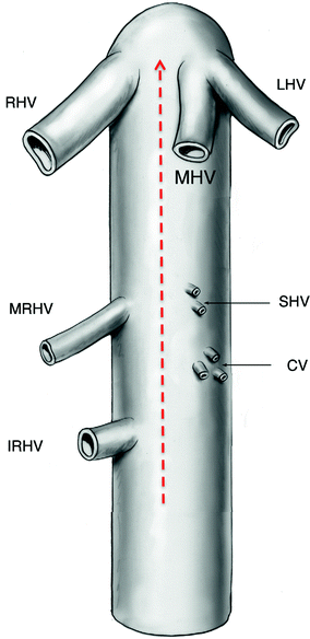
Fig. 1.2
Direction and position for the clamp during Belghiti’s liver-hanging maneuver. RHV right hepatic vein; LHV left hepatic vein; MHV middle hepatic vein; SHV short hepatic vein; CV caudate vein; MRHV middle right hepatic vein; IRHV inferior right hepatic vein. Reprinted from Am J Surg 193; Gaujoux S, Douard R, Ettorre GM, Delmas V, Chevallier J-M, Cugnenc P-H. Liver-hanging maneuver: an anatomic and a clinical review. p.488–492; copyright © 2007; with permission from Elsevier
Belghiti’s LHM uses a tape to pass through the retrohepatic tunnel between the anterior surface of the inferior vena cava (IVC) and the liver parenchyma. This technique has been found useful in selected patients who undergo left or right hepatectomy. The key to this maneuver is the blind dissection of the retrohepatic space anterior to the IVC using a long vascular clamp. After blind dissection starting inferiorly, the clamp appears between the right and the middle hepatic veins. A tape is seized with the clamp and is passed through the retrohepatic tunnel. During parenchymal division, upward traction on this tape around the liver contributes to better exposure and hemostasis of the deeper hepatic parenchyma in front of the IVC. Many surgeons are still reluctant to use this maneuver. Some consider it too complicated and difficult, whereas others are afraid of bleeding from an injured short hepatic vein during retrohepatic dissection, the reported incidence of which is about 4–6% [4–6].
To overcome these difficulties, a modification of this technique—the liver double-hanging maneuver—was devised. This technique has the advantage of being technically simple and safe.
Chen’s Double-Hanging Maneuver
The liver is exposed through a right subcostal incision with midline extension, which can be extended to the left side for better exposure if necessary. The ligamentum teres and the falciform ligament are dissected. A tape is placed around the duodenohepatic ligament and the infrahepatic IVC, respectively (Fig. 1.3). These tapes can be used to control hemorrhage if necessary. The peritoneum on the right side of the IVC just inferior to the liver is open to expose the right adrenal gland. The operator then uses his/her right index finger to dissect the space from below upward between the hepatic parenchyma and the anterior and superior edge of the right adrenal gland, and then along the right side of the IVC.
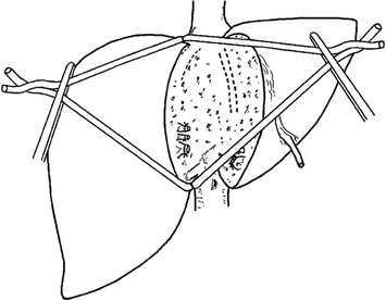
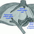
Fig. 1.3
Liver double-hanging maneuver. Reprinted from Surgery 144; from Chen XP, Zhang WG, Lau WY, Qiu FZ. Right hepactectomy using the liver double-hanging maneuver through the retrohepatic avascular tunnel on the right of the inferior vena cava. p. 830–833; copyright © 2008; with permission from Elsevier
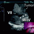
The right coronary ligament is open suprahepatically on the right side of the IVC for 2–3 cm. The operator uses his/her left index finger to dissect the retrohepatic space from above downward along the right side of the IVC. The retrohepatic tunnel is built when the fingers touch each other. A kidney pedicle forceps is used to place two tapes around the liver for liver suspension. One tape is pulled to the right, and the other is pulled to the left. Liver transection is performed along a plane to the right of the middle hepatic vein as determined by intraoperative ultrasound. The liver double-hanging maneuver contributes to better exposure of the operative fields and is easier to manipulate during liver parenchymal transection (Fig. 1.4). When the liver parenchymal transection reaches to the right hepatic vein, the origin of the right hepatic vein from the IVC is dissected, doubly ligated, and divided. Liver transection is then carried out along the right border of the IVC, which divides the caudate process and then ligates and divides the short hepatic veins.
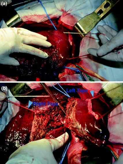
Fig. 1.4
Chen’s liver double-hanging maneuver during surgery. The two-hanging tapes were used to guide the transection plane, control bleeding, minimize tumor touch, and protect the IVC. Figure A is reprinted from Surgery 144; Chen XP, Zhang WG, Lau WY, Qiu FZ. Right hepactectomy using the liver double-hanging maneuver through the retrohepatic avascular tunnel on the right of the inferior vena cava. p. 830–833; copyright © 2008; with permission from Elsevier
In this improved technique, the tunnel goes through a true avascular space that contains loose connective tissues only. The right adrenal grand is an important anatomic structure in this retrohepatic space. If it is not injured, then the risk of bleeding is very low. During the dissection of the retrohepatic tunnel, the operator should keep his or her index finger close to the back of the liver. This maneuver is simple and safe because blind dissection of the space between the right and middle hepatic veins is not needed.
In this technique of right hepatectomy, the entire caudate lobe is left intact, whereas in Belghiti’s liver-hanging technique, parts of the paracaval portion and the caudate process are resected. Although the difference in the amount of liver tissue resected between these two techniques is minimal, Chen’s technique represents a right hepatectomy, which is more correct anatomically.
In conclusion, the liver double-hanging maneuver for major right hepatectomy has several advantages: First, the operator can feel the retrohepatic tissues with the index fingers, which is safer than blind dissection using forceps. Second, it is not necessary to dissect between the right and the middle hepatic veins. Third, a true avascular space that contains loose connective tissue exists only behind the liver parenchyma on the right side of the IVC. The success rate of tunneling through this retrohepatic space is 100%. Fourth, the leftward and rightward traction on the tapes contributes to better exposure of the deeper parenchymal tissue during liver transection. Tightening the tapes helps to control bleeding from the hepatic transection plane, especially for bleeding coming from branches of the hepatic veins. In conclusion, the liver double-hanging maneuver through the retrohepatic tunnel on the right side of the IVC is a safe and easy procedure.
Modified double-hanging maneuver, namely single-hanging tape on the remnant side, may be applied to guide the transection plane during right hemi-hepatectomy when the liver can be exposed well (Fig. 1.5). When the abdominal cavity has enough space to expose the liver, or when the right liver lobe could be partially mobilized, the right tape is not necessary in some cases. In this condition, the single left tape is used to guide the transection plane and protect the IVC. This single-hanging maneuver through the retrohepatic avascular tunnel on the right of the IVC has also been applied successfully to laparoscopic or robotic-assisted laparoscopic right hemi-hepatectomy in more than 100 cases in our center (Fig. 1.5).
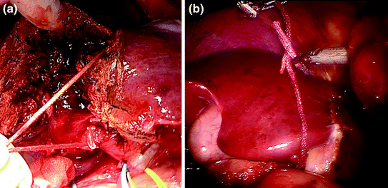
Fig. 1.5
Modified double-hanging maneuver. a Single-hanging tape on the remnant side was applied to guide the transection plane and protect the IVC. b The single-hanging maneuver through the retrohepatic avascular tunnel on the right of the IVC was applied to robotic-assisted laparoscopic right hemi-hepatectomy
Case Presentation
A 68-year-old woman, who is a hepatitis B virus (HBV) carrier, claimed abdominal distension, without jaundice or fever. No family or genetic history was found. She had hepatitis B virus infection, but had not accepted any therapy before admission. Neither history of cigarette smoking nor alcohol drinking was found. Preoperative computed tomography (CT) revealed huge HCC in the right lobe of the liver, with suspected diaphragm invasion. Neither intrahepatic metastasis nor lymphonode metastasis was shown in CT scanning. It also showed suspected tumor thrombus in the right branch of the portal vein, but tumor pressure of the portal vein could not be excluded (Fig. 1.6). However, ultrasonography did not show portal vein tumor thrombus. Preoperative biochemical examinations revealed AFP: 8760 ng/ml, HBV-DNA 3.43 × 10³ copies/ml and Child-Pugh A grade.
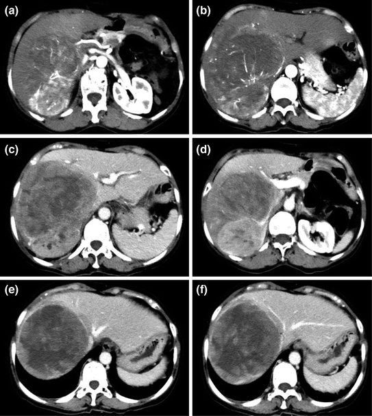
Fig. 1.6
CT scanning for the patient with HCC located in the right lobe. The arterial phase showed identical HCC enhancement (a) and suspected diaphragm invasion (b), but no lymphonode metastasis was presented; c, d the portal vein phase showed normal left branch of the portal vein, and suspected tumor pressure or tumor thrombus in the right branch of the portal vein; e, f the delayed phase showed the common branch of middle hepatic vein and left hepatic vein (e), and indicated that the middle hepatic vein was pressed by the tumor (f). The right hepatic vein could not be distinguished, probably was circled or pressed by the tumor
Our Management
- 1.
Evaluation of the Remnant Liver FunctionRelated posts:
 Resection of Centrally Located Cystadenoma/Cystadenocarcinoma
 Debulking of Extensive Neuroendocrine Liver Metastases
 Management of the Gangrenous Gallbladder
 Totally Laparoscopic Right Hepatectomy Combined with En-Bloc Partial Resection of the Inferior Vena Cava
 Gallbladder Cancer with Common Bile Duct Invasion
 Resection of Renal Cell Carcinoma Involving the Liver with Tumor Thrombus Extending into Inferior Vena Cava Requiring Venovenous Bypass

Stay updated, free articles. Join our Telegram channel

Full access? Get Clinical Tree


