Fig. 3.1
Imaging studies of the first case that exhibit a symptomatic centrally located cystic lesion with enhancing intramural projections. The cystic lesion abuts the left hepatic duct, producing significant left hepatic duct dilation (a–d). No vascular invasion was apparent. In e, the postoperative imaging after extended left hepatectomy. The lesion proved to be an intrahepatic cystadenocarcinoma
Procedure
Based on the imaging and, in particular, the recent changes observed, invasive malignancy was strongly suspected; accordingly, extended left and caudate hepatectomy (segments 1, 2, 3, 4, and 8) was performed. The patient’s anatomy was such that the segment 5 pedicle arose from the “posterior” right pedicle, facilitating salvage of segment 5. The left hepatic artery and portal vein were dissected and ligated extrahepatically, and the left hepatic duct was transected close to the bifurcation and oversewn. The caudate was elevated off of the cava after dividing short hepatic veins. The left and middle hepatic veins were encircled above the liver and taken flush with the cava using a stapling device. Anteriorly, the parenchymal division began through liver parenchyma away from the cyst, using an electrosurgical device without hilar occlusion, between segments 4 and 5; the dissection subsequently proceeded to the right, following the contour of the tumor, up along the right anterior portal structures in an extra-Glissonian plane, and the segment 8 pedicle was taken within the parenchyma. Dorsally, the cyst/tumor extended rightward to the right hepatic vein; dissection was carried out in the plane of the vein and the tumor was successfully separated away except for a small point of adherence, whereupon a side-biting vascular clamp was applied onto the vein, a portion of the vein wall was excised, and repair was carried out with 6-0 polypropylene.
Outcome
The postoperative course was uneventful, and the patient was discharged on postoperative day 4. Pathology showed moderately to poorly differentiated IBCC with extensive lymphovascular and perineural invasion arising in an IBC; margins and lymph nodes (0/2) were free of tumor. She received adjuvant gemcitabine for 6 months, and was alive and free of recurrence on followup 3 years after surgery.
Case 2
History
A 52-year-old male with no significant past medical history presented with upper abdominal fullness and abnormal liver tests, with alkaline phosphatase 492, ALT 163, and bilirubin 1.4. CEA and CA19-9 were normal. MR showed a 20 cm multiloculated cyst involving segments 4/5/8 and compressing but not invading the left and right anterior portal structures (Fig. 3.2a–d). There was no evidence of portal hypertension.
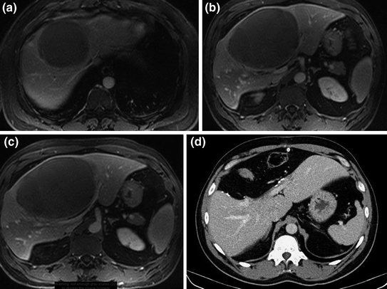
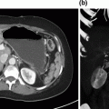
Fig. 3.2
Study imaging of the second case presenting with asymptomatic centrally located cystic lesion (a–c). There are no intracystic projections and no vascular involvement. The intrahepatic biliary tree is not dilated. The patient underwent a central pericystectomy (d). The lesion proved to be an intrahepatic cystadenoma with foci of cystadenocarcinoma
Procedure
Based on the smooth, well-defined border of the cyst with compression but with no evidence of invasion of portal structures, central resection in the enucleation plane of the lesion was undertaken. After taking down the gallbladder, tying and dividing the gallbladder mesentery, and lowering the portal bifurcation in an extra-Glissonian plane, the segment 4 structures were dissected and ligated in the umbilical fissure. The capsule of the liver was scored with electrocautery around the edge of the cyst. The porta hepatis was occluded en masse with a broad vascular clamp for 19 min, during which the cyst was separated from segments 2–3 in the enucleation plane using scissors dissection, clipping vessels, and ducts that traversed the transection plane with a multiclip applier. After a 5-minute period of reperfusion, the porta was once again clamped for 20 min, during which the cyst was similarly enucleated away from segments 5–8. The main trunk of the middle hepatic vein, which was closely applied onto the cyst, was ligated at its junction with the left hepatic vein and removed along with the specimen.
Outcome
The patient had an uneventful recovery and was discharged home on postoperative day 4. Pathology revealed an 18 cm hepatobiliary mucinous cystadenoma with areas of carcinoma; extensive necrosis, cystic degeneration, and limited lymphovascular invasion were noted. No adjuvant therapy was given. The patient was alive and free of recurrence at 2 years after surgery.
Technical Pearls
Dissection of the portal structures in the extraglissonian plane by lowering the hilar plate is the most expedient and safest approach.
If complete right anterior resection is planned the right anterior sectoral structures can be encircled and divided using the ultrasonic dissector.
Dissection and encircling of the middle hepatic vein above the liver can generally be accomplished, and is facilitated by making a short incision into the liver parenchyma overlying the confluence.
A scissors dissection technique, using the blunt scissors tip to dissect through parenchyma and identify small vascular structures, which can be clipped, can be used for parenchymal transection.
Discussion
The central portion of the liver comprises segments 4, 5, and 8; depending on the nature, size, and location of the pathology to be dealt with, resection of centrally located tumors may require removal of part or all of one or more of these segments (Fig. 3.3). Central resection requires division of the liver twice, with two resultant cut surfaces and the attendant risks. Extended right or left hepatectomy is an alternative that has, over the years, been commonly employed because of its relative technical simplicity; central resection has become more common of late with the recognition of the value of parenchyma-sparing surgery, both for primary liver tumors where there is concern about hepatic functional reserve, and for metastatic tumors where it is desirable to maintain options to treat possible future recurrence [9].
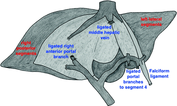
Fig. 3.3
Schematic representation of a central hepatectomy (removal of segments 4, 5, 8)
Anatomical Considerations
Segment 4 is commonly described as having two subsegments, 4A and 4B, although there are typically multiple portal branches to segment 4 rather than two discrete pedicles. Be that as it may, the left portal vein runs transversely to the umbilical fissure under segment 4B and then up the umbilical fissure, terminating in the obliterated umbilical vein, often designated as the round ligament. The segment 4 portal branches, along with those from the hepatic artery and hepatic duct, enter segment 4 in the umbilical fissure and course through segment 4 from left to right (see Fig. 3.3). These portal structures are readily clipped or ligated in the umbilical fissure when complete resection of segment 4 or 4B is planned, but the fact that they run from left to right makes it possible to divide through segment 4 at any distance from the umbilical fissure without preliminary dissection while maintaining perfusion and biliary drainage to the portion of segment 4 that is left behind [10]. As they approach the liver, the portal vein, hepatic artery, and bile duct are invested and subsequently distributed through the liver within a common sheath derived from the reflection of Glisson’s capsule, and in many circumstances dissection of these structures in the extra-Glissonian plane is both the most expedient and the safest approach [10]. Separating the portal structures away from the parenchyma of segments 4B and 5 is a key maneuver when complete resection of these segments is planned. This so-called lowering of the hilar plate is accomplished by first removing the gallbladder if present, ligating and dividing the tissue at the base of the gallbladder fossa (commonly called the gallbladder mesentery; there are often small structures there that are of no consequence, but that can be troublesome if not ligated), dividing the peritoneum under segments 4B and 5 as it envelopes the portal structures, and separating the portal structures from the liver parenchyma outside of Glisson’s sheath. It typically requires the division of a few small portal branches near the portal bifurcation to establish this extra-Glissonian plane. While this process of lowering the hilar plate can be carried out with simple scissors dissection, an ultrasonic dissector, when available, is a useful tool to establish and maintain the correct plane of dissection.
Related posts:
 Debulking of Extensive Neuroendocrine Liver Metastases
 Multifocal Branch-Duct Intraductal Papillary Mucinous Neoplasm
 Pancreatic Adenocarcinoma in the Neck of the Pancreas Involving the Celiac Trunk (Appleby Procedure)
 Gallbladder Cancer with Common Bile Duct Invasion
 Chronic Pancreatitis: Puestow and Frey Procedures
 Resection of Renal Cell Carcinoma Involving the Liver with Tumor Thrombus Extending into Inferior Vena Cava Requiring Venovenous Bypass
Stay updated, free articles. Join our Telegram channel

Full access? Get Clinical Tree


