Reproductive Disorders


Reproductive Disorders


Neil Goodman

Rhoda H. Cobin


AMENORRHEA


Etiology

There is considerable overlap in the causes of primary and secondary amenorrhea since some disorders may begin either before or after puberty and the onset of menses. Ovarian dysgenesis and müllerian defects, including uterine and outlet disorders, account for about 60% of patients with primary amenorrhea. Hyperandrogenic disorders, primary ovarian insufficiency, and hypothalamic amenorrhea account for most patients with secondary amenorrhea. Pregnancy should always be considered as a diagnosis. Constitutional delay of puberty is often responsible for what would otherwise appear to be primary amenorrhea.


Epidemiology

The incidence of primary amenorrhea varies from 0.48% to 1.2%. That of secondary amenorrhea is about 4.9%.


Pathophysiology

The approach to amenorrhea depends on whether it is primary or secondary. In the former, defects may be present at any level of the reproductive system (i.e., hypothalamus, pituitary, ovaries, uterus, or vaginal outflow tract). In women who have had prior menses, it is clear that not only must the woman have had an anatomically normal uterus and outflow tract but that prior normal stimulation with estrogen has occurred, implying ovarian function.


Pregnancy should always be excluded in any patient first seen with amenorrhea, particularly secondary amenorrhea or primary amenorrhea with normal development. Thyroid abnormalities are commonly associated with menstrual disorders and should be sought with an initial thyroid-stimulating hormone (TSH) level.

A history and physical examination will help to define the problem and suggest appropriate laboratory investigation.


Primary Amenorrhea


Vaginal, Uterine, and Ovarian Disorders

Imperforate hymen will be seen with normal growth and development, normal secondary sexual characteristics, and, often, premenstrual molimina and lower abdominal cramping at the time of expected menses. The diagnosis is made by physical examination, and the treatment is surgical hymenectomy.

Müllerian agenesis (Mayer-Rokitansky syndrome) occurs with normal growth and development of secondary sexual characteristics. A rudimentary vaginal canal may be present but with absent uterus and fallopian tubes. Associated abnormalities (including scoliosis, unilateral renal agenesis, and, rarely, cardiac defects) may be found [1]. Although the disorder is usually sporadic, various genetic etiologies have been proposed [2, 3]. The diagnosis is made with sonography. Levels of estrogen and gonadotropins are normal. Treatment may include vaginal reconstructive procedures.

Failure of normal ovarian development may be due to Turner syndrome (TS), which may be clinically suspected by the observation of undeveloped secondary sexual characteristics, short stature, and somatic abnormalities including widely spaced nipples, low-set hairline, and other skeletal abnormalities. The diagnosis of an ovarian etiology is confirmed with the finding of an elevated follicle-stimulating hormone (FSH) due to lack of ovarian inhibin production. XO is the most frequent genetic abnormality, but other X chromosome abnormalities and mosaicism may be present. Variants with Y chromosomal material may have mild virilization. The presence of Y chromosome increases the risk of gonadoblastomas, and thus, gonadectomy should be considered [4].

Primary ovarian dysgenesis with XX karyotype is a rare condition that may be inherited as an autosomal recessive trait and does not have the associated somatic features of TS; it may be associated with tall stature. Failure of development of secondary sexual characteristics, ovarian dysgenesis, and normal uterine and vaginal structures are noted. FSH is elevated [5, 6].

Rarely, autoimmune oophoritis will be seen as primary amenorrhea, although it is more likely to occur as secondary amenorrhea with evidence of ovarian failure (i.e., elevated FSH). Antiovarian antibodies are sometimes detectable, and a strong association exists with other autoimmune diseases.

Androgen insensitivity syndrome results from mutations in the androgen receptor of varying types and severity with corresponding variation in clinical presentation, including phenotypic females with normal breast tissue, modest axillary and pubic hair, and absent müllerian structures. The incidence is higher in females with inguinal hernias [7, 8]. The diagnosis is made with sonography and the finding of a male-range testosterone. The karyotype is XY.


Pituitary/Hypothalamic Disease

Hypogonadotropic hypogonadism (low luteinizing hormone [LH] and FSH) may be seen as primary amenorrhea with or without anosmia (Kallmann syndrome). Various mutations in the KAL gene, which cause abnormal migration of gonadotropin-releasing hormone (GnRH)-producing cells to the hypothalamus have
been described [9]. Other less common syndromes first seen as primary hypogonadotropic hypogonadism include Prader-Willi syndrome and Laurence-Moon-Biedel syndrome.

Pituitary and hypothalamic tumors, especially craniopharyngioma, as well as infiltrative diseases, may rarely begin in childhood presenting as primary amenorrhea, although they usually begin later in life and therefore after menses have begun (vide infra).


Secondary Amenorrhea


Vaginal/Uterine Disorders

In patients with previously normal menses, secondary amenorrhea with normal hormonal function and abnormal uterine function may develop. A history of pelvic infection, dilation and curettage, or uterine instrumentation in a patient with secondary amenorrhea should suggest Asherman syndrome. The absence of bleeding after estrogen/progesterone therapy implies that a functional endometrium is absent. Hysteroscopy or hysterosalpingography may be required to establish the diagnosis.


Ovarian Failure

Autoimmune oophoritis will occur as secondary amenorrhea with evidence of ovarian failure (i.e., elevated FSH). Antiovarian antibodies are sometimes detectable, and a strong association with other autoimmune diseases is present [10, 11]. Premature ovarian failure is a feature of the fragile X syndrome. Genetic analysis and counseling are required. Premature ovarian failure may be familial and may be found with no evidence of autoimmune disease.


Pituitary and Hypothalamic Disease

Women with normal or low gonadotropins, normal pelvic structures, and normal androgens may be suspected of having hypothalamic or pituitary disease. Although the latter is often functional, it is imperative to image the pituitary with CT or MRI to rule out pituitary tumors or other lesions in this area, which may affect normal hypothalamic—pituitary function. Usually, this is seen as secondary amenorrhea, although occasionally, the onset is early enough in life to appear as primary amenorrhea.

A high prolactin level may indicate either prolactin production from a pituitary adenoma or stalk compression, causing reduced dopaminergic negative regulation of pituitary prolactin production. Because the level of prolactin is usually proportionate to the size of the tumor in pure prolactin-secreting tumors, tumors disproportionately large compared with the serum prolactin level should be suspected of being nonprolactinomas, which are not amenable to therapy with dopaminergic agents and may be considered for surgery.

Hypothalamic amenorrhea is a very common cause of secondary amenorrhea and an occasional cause of primary amenorrhea. Laboratory studies reveal normal to low gonadotropins and estrogen and normal androgens and prolactin levels. Anatomic pathology is absent. A thorough and sensitive history must be obtained because emotional stress, excessive exercising, weight loss, dieting, and eating disorders such as anorexia and bulimia may be difficult to elicit on initial history. Once rapport is established, the patient may be more comfortable in discussing these important etiologic factors and their management.


Hyperandrogenism

Excessive androgen production is associated with both primary and secondary amenorrhea and may be due to either ovarian or adrenal sources.

The most common cause of primary amenorrhea with excess androgen production is adrenal hyperplasia, most frequently 21-hydroxylase deficiency. The
frequency of this disorder is variable, depending on ethnic origin. Presentation may be at birth with ambiguous genitalia, or it may be delayed until later in life, particularly in the non—salt-wasting variety. In childhood, accelerated growth and bone maturation and signs of hyperandrogenism (hirsutism, acne, increased musculature, alopecia) appear. If untreated, amenorrhea and short final adult stature are found (because of early closure of the epiphyses secondary to androgenic stimulation). The diagnosis is made with the finding of elevated 17-OH progesterone, either in the basal state or with adrenocorticotropic hormone (ACTH) stimulation. Genetic testing will confirm the presence of a variety of mutations of the CYP21 gene on chromosome 6 [12]. Treatment consists of glucocorticoid replacement to suppress excess ACTH and hence adrenal androgen production, with care to avoid excess glucocorticoid, which can cause growth retardation, osteopenia, and iatrogenic Cushing disease [13, 14].

Other less common enzyme defects causing adrenal hyperplasia include: 11-hydroxylase deficiency, which occurs with amenorrhea, hyperandrogenism, hypertension, and hypokalemia; 3β-hydroxysteroid dehydrogenase deficiency, which causes hyperandrogenism; and 17-hydroxylase deficiency, which occurs with sexual infantilism.

Polycystic ovarian syndrome (PCOS) affects between 6% and 10% of women of reproductive age and typically occurs with oligomenorrhea dating from the onset of puberty, along with variable hyperandrogenism. PCOS is strongly associated with insulin resistance and carries a high risk of glucose intolerance, frank diabetes, hypertension, dyslipidemia, and an increased frequency of myocardial infarction and cerebrovascular disease in later life; making a diagnosis is critical [15].

Signs or symptoms of hyperandrogenism may include acne, hirsutism, and alopecia. More severe hyperandrogenism (virilization) including increased muscle mass, clitoromegaly, deepening of the voice, and male-pattern baldness is indicative of higher testosterone levels (vide infra), usually not seen with PCOS.


HYPERANDROGENISM AND PCOS


Hyperandrogenism

Hyperandrogenism describes the reaction of the pilosebaceous units of the skin when stimulated by androgens. The skin reacts by excessive hair growth in a
male pattern distribution (hirsutism), acne, or hair loss (alopecia). However, the finding of hyperandrogenism does not define excess androgen production but can be a significant indicator of the presence of an androgen excess disorder, leading to the measurement of testosterone and other androgens, evaluating if there is an overproduction by the ovaries or adrenal glands. Hirsutism may occur without actual elevation in androgen levels if increased target organ sensitivity to androgen action exists. It should be noted that ethnic differences in the number of hair follicles present and individual skin sensitivity of the pilosebaceous unit to androgens are major determinants of the presence of hirsutism, as well as acne and androgenic alopecia [38].


Table 5.1. Amenorrhea Summary


Uterus


FSH


Prolactin


Testosterone


Karyotype


Treatment


Primary Amenorrhea


Turner syndrome


Yes


N


N


45,X; mosaic variants


Counseling; HRT consideration; CV evaluation; thyroid tests; GH? and discussion if appropriate


Immune ovarian failure


Yes


N


N


46,XX


HRT?


Gonadal dysgenesis


Yes


N


N


46,XX; 46,XY


remove gonads if Y or part of Y chromosome present


21α-Hydroxylase deficiency


Yes


N


N


46,XX


Glucocorticoids


17α-Hydroxylase deficiency


Yes


N


N


N


46,XX


Glucocorticoids; HRT


Androgen insensitivity


Yes


N


N


46,XY


Remove gonads


Müllerian agenesis


No


N


N


N


46,XX


Surgical consideration; evaluate for urinary tract anomalies


Kallmann syndrome


Yes


N,↓


N


N


46,XX


hMG/GnRH for fertility


Hypothalamic dysfunction


Yes


N,↓


N


N


46,XX


hMG/GnRH for fertility if primary disease not treatable


Prolactinoma


Yes


N


N


46,XX


Dopamine agonists, surgery


Other pituitary tumor


Yes


N,↓


N,↑


N


46,XX


Surgery, irradiation


Infiltrative disorders


Yes


N,↓


N,↑


N


46,XX


Treat disease


Secondary Amenorrhea


Premature ovarian failure


Yes


N


N


46,XX


HRT


Iatrogenic: radiotherapy, chemotherapy


Yes


N


N


46,XX


HRT


Low body weight/exercise


Yes


N,↓


N


N


46,XX


Weight gain as appropriate


Hypothalamic dysfunction


Yes


N,↓


N,↑


N


46,XX


HRT, hMG/GnRH if cannot treat underlying disease for fertility


Sheehan syndrome


Yes


N


N


N


46,XX


HRT, hMG/GnRH if cannot treat underlying disease for fertility


Prolactinoma


Yes


N


N


46,XX


Dopamine agonists, surgery; hMG if hypopituitary for fertility


Hyperprolactinemia


Yes


N,↓


N


46,XX


Change of medications possible? Treat underlying medical problem


Other pituitary tumor


Yes


N,↓


N,↑


N


46,XX


Surgery, irradiation


Infiltrative disorders


Yes


N


N,↑


N


46,XX


Treat disease


PCOS


Yes


N


N,↑


N,↓


46,XX


See section on weight control; metformin; OCT


Asherman syndrome


Yes


N


N


N


46,XX


Surgery if appropriate


GH, growth hormone; GnRH, gonadotropin-releasing hormone; hMG, human menopausal gonadotropin; HRT, hormone replacement therapy; N, normal; PCOS, polycystic ovarian syndrome.


Hirsutism should not be confused with hypertrichosis, which is defined as the growth of hair on any part of the body, in excess of the amount usually present in persons of the same age, race, and sex. Extreme excess of androgen production will present as virilization, in which hirsutism is accompanied by male-pattern baldness, deepening of the voice, increased muscle development, and clitoromegaly. The presence of true virilization usually points to a serious underlying disorder, such as virilizing tumors of the ovary or adrenal gland or hyperthecosis ovarii [39].

Girls with severe acne that is persistent or exacerbates in the mid-20s or 30s or that is resistant to oral and topical agents have a high incidence of hyperandrogenemia and a 40% likelihood of having PCOS [40, 41]. Androgenic alopecia typically presents as thinning of the anterior midline area of the scalp and extends posteriorly to the midvertex of the scalp. The frontal hair line is usually preserved, and a triangular pattern of hair thinning (triangle sign) is often noted in the anterior midline area. Alopecia is present in 40% to 70% of women with PCOS [42].


PCOS

The most common form of hyperandrogenism is the polycystic ovary syndrome (PCOS). This heterogeneous syndrome is the most frequently encountered endocrine disturbance in women of reproductive age. The prevalence of PCOS ranges from 5% to 10% [43], and it affects all ethnic groups. PCOS is not only a reproductive disorder but also a metabolic one [44].

Defining the syndrome includes four issues: oligomenorrhea, clinical and biochemical features of hyperandrogenism, polycystic ovaries by ultrasonography, and the exclusion of other causes associated with androgen excess (nonclassical congenital adrenal hyperplasia (NCCAH), virilizing syndromes of the ovary or adrenal, and Cushing syndrome). The consensus is that oligomenorrhea and hyperandrogenism are essential components of the syndrome [45]. The specificity of morphologic evidence of polycystic ovaries on ultrasonography is limited since 23% of apparently normal women have characteristic findings of polycystic ovaries [46].

Disruption of the female reproductive system in PCOS typically begins at menarche, with menstrual cycles longer than 35 to 40 days or fewer than 10 periods per year, defined as oligomenorrhea. The irregular menses may be interspersed with episodes of what appear to be regular cycles (not necessarily ovulatory) and may also be associated with episodes of heavy dysfunctional intermenstrual bleeding [45].


PCOS AND INSULIN RESISTANCE (METABOLIC SYNDROME)

The pathophysiology of the menstrual irregularities and hyperandrogenism of PCOS includes hypothalamic—pituitary abnormalities that result in GnRH and LH dysfunction and a primary enzymatic defect in ovarian or combined ovarian and adrenal steroidogenesis. Observational studies of obese women with PCOS have found an increased risk for developing impaired glucose tolerance (IGT)
(31%—35%) and type 2 diabetes mellitus (DM) (7.5%—10%) [47]. With 50% to 70% of all women with PCOS having some degree of insulin resistance [48], a hypothesis to explain these aspects of PCOS therefore follows that this metabolic disorder of compensatory hyperinsulinemia exerts adverse effects on the hypothalamus, pituitary, ovaries, and, possibly, adrenal glands, explaining the oligomenorrhea and hyperandrogenemia.

There are serious health consequences beyond dermatologic and reproductive dysfunction secondary to the insulin resistance, which are defined by the metabolic syndrome [49]. The metabolic syndrome includes:


  • Glucose intolerance/diabetes


  • Elevated blood pressure level (≥130/85)


  • Increased waist circumference (≥35 inches or waist-to-hip)


  • Reduced high-density lipoprotein cholesterol level (≤50 mg/dl)


  • Elevated triglyceride levels (≥150 mg/dl)

All women with PCOS should be screened with a 2-hour (75-g) glucose tolerance test [50], with rescreening every 1 to 2 years because of the high rates of conversion to IGT and diabetes. Of clinical note is that acanthosis nigricans with or without skin tags are markers of hyperinsulinism and the MS [51].


PRECOCIOUS PUBERTY


Pathophysiology

Central precocious puberty (CPP) is defined as early activation of pulsatile GnRH activity in an otherwise normal hypothalamopituitary—gonadal axis. Pseudopuberty occurs when true central activation is absent.


Etiology and Diagnosis

Premature adrenarche and thelarche are characterized by prepubertal LH, FSH, testosterone, and estradiol levels, without progression of bone age. In premature adrenarche, a significantly elevated DHEA-S level requires further evaluation for CAH or adrenal tumor. Girls with premature adrenarche may be at risk for PCOS and insulin resistance. Periodic monitoring for both conditions is indicated to ensure they do not progress.

In girls, CPP is overwhelmingly idiopathic but may result from a hypothalamic hamartoma with pulsatile GnRH secretion. Bone-age testing should be undertaken to assess the effect on growth rate and to gauge progression and response to therapy. MRI of the brain/pituitary is needed in all cases of CPP to exclude disease affecting the central nervous system. In boys, CPP is nearly always secondary to a tumor, irradiation, or septooptic dysplasia with premature activation
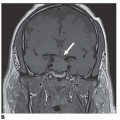
of GnRH secretion. Discontinuation after exposure to exogenous sex steroids may also result in activation of the central axis, resulting in acquired CPP. MRI of the brain and hypothalamopituitary region should be done, as well as a bone-age test.

Pseudopuberty in girls is associated with prepubertal levels of LH and FSH, LH unresponsive to GnRH stimulation, and sex hormones in the pubertal range or higher. Causes in girls include ovarian follicular cysts, McCune-Albright syndrome (associated with café-au-lait spots and polyostotic fibrous dysplasia), and stromal cell tumors of the ovary. A pelvic ultrasound should be done to look for cysts or masses in the ovary. Thyroid studies should be done to evaluate for hypothyroidism.

In boys, autonomous testicular function may be present secondary to a Leydig cell tumor, human chorionic gonadotropin (hCG)-secreting tumor, McCune-Albright syndrome (activated G protein), or familial male precocious puberty caused by an activating mutation of the LH receptor expressed in testicular Leydig cells. LH and FSH levels will be in the prepubertal range, with pubertal range or higher testosterone levels. With a Leydig cell tumor, a testicular mass may be noted on examination or ultrasound. An hCG level should be checked in boys with precocious puberty because tumors that secrete hCG can stimulate testicular testosterone production. These tumors may occur in the gonads, pineal, liver, posterior mediastinum, or retroperitoneum.

Other causes of pseudopuberty include abuse of exogenous androgens, congenital adrenal hyperplasia (CAH), and a pituitary gonadotroph-secreting adenoma. Thyroid studies (TSH, FT4) should be done to evaluate for hypothyroidism.

Aug 2, 2016 | Posted by in ENDOCRINOLOGY | Comments Off on Reproductive Disorders

Full access? Get Clinical Tree

Get Clinical Tree app for offline access