Reactions in Patients with Skin of Color
Oyetewa Oyerinde
Michelle S. Lee
Jennifer Y. Lin
As is the case for the full spectrum of dermatologic disease, manifestations of cutaneous toxicity from anticancer therapy differ across skin types. Better recognition of these toxicities in skin of color can help decrease potential racial disparities in clinical outcomes in oncology.
When we consider skin color, we consider both intrinsic pigment and facultative pigment (pigment acquired in response to outside stimulus). Facultative pigmentation is most commonly seen in the setting of ultraviolet radiation (UVR) in the form of a tan. Chemotherapeutics that are DNA-damaging or alkylating often mimic UVR and can lead to melanin release. Thus, patients with darker skin types or those who tend to tan easily are at increased risk of hyperpigmentation from certain anticancer therapies.
Inflammatory conditions can also release or disrupt melanin transfer in keratinocytes resulting in postinflammatory hyperpigmentation or hypopigmentation. Postinflammatory hyperpigmentation occurs with greater frequency and persists for longer periods of time in darker skin. In the case of postinflammatory hypopigmentation, the contrast between the normal skin and affected skin is greater, and thus more prominent in darker skin types, often leading to additional and prolonged distress in these patients; the impact of dyspigmentation on quality of life and anticancer therapy adherence warrant appropriate attention.
Conventional management of patients experiencing pigmentary complications includes treating the underlying cause of inflammation and waiting for the inflammation to resolve before treating the resultant pigmentary change. There are several modalities now available to hasten the resolution of hyperpigmentation and hypopigmentation (Table 1.1). Interventional treatments should be administered by experienced practitioners, as several of these modalities can potentially worsen hyper/hypopigmentation.
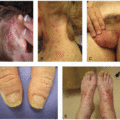
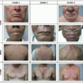
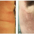
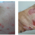
For many practitioners, it can be difficult to discern subtle pigmentary changes and/or erythema in dark skin tones. Inflammatory conditions can look dark pink to violaceous in darker skinned patients (Figures 1.1 and 1.2). Always asking about symptomatology, such as itch or burning sensation, becomes even more important in assessing the type of toxicity the patient may be experiencing.
There is a paucity of literature related to disparities in the diagnosis and treatment of cancer therapy-related cutaneous toxicities across skin type and ethnic groups. Existing
literature regarding these reactions in skin of color is limited mostly to clinical trials in Asia, which describe incidence rates of skin toxicities in Japan, China, and South Korea.7,8,9,10,11,12,13,14,15,16,17,18
literature regarding these reactions in skin of color is limited mostly to clinical trials in Asia, which describe incidence rates of skin toxicities in Japan, China, and South Korea.7,8,9,10,11,12,13,14,15,16,17,18
TABLE 1.1 • Prophylaxis and Treatment Options for Hypo- and Hyperpigmentation in Skin of Color
Stay updated, free articles. Join our Telegram channel
Full access? Get Clinical Tree
 Get Clinical Tree app for offline access

|
|---|

