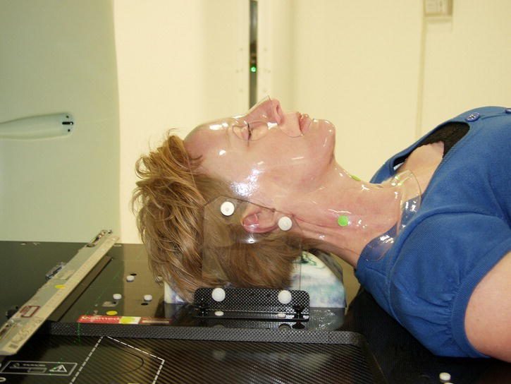
Radiotherapy side effects andtheir management

Chapter 5
Radiotherapy side effects andtheir management


Alexandra Gilbert, Emma Dugdale and Robin Prestwich


St James’ Institute of Oncology, UK


Overview of radiotherapy toxicity


Radiotherapy itself is painless during delivery. Radiotherapy is a localised treatment and therefore other than treatment-related fatigue, side effects are related to the anatomical area of the body that is receiving treatment. For example treatment to the thorax will not cause lower GI symptoms such as diarrhoea.


There are a number of clinician reported acute and late toxicity grading systems in clinical use, including the Common Terminology Criteria for Adverse Events (CTCAE version 4).


There are three types of toxicity:


  1. Acute toxicity: for example mucositis

    1. Develops during treatment, usually after the first 5–10 fractions and peaks at 2–4 weeks post completion.
    2. Increasesrcinogenic and there is a risk during treatment and is maximal in the first few weeks following the end of treatment.
    3. Generally reversible but must be managed appropriately to ensure patient compliance with full course of treatment.

  2. Delayed toxicity: for example radiation pneumonitis. develops at least six weeks after completion of radiotherapy and must be recognised as such to ensure it is appropriately managed.
  3. Late toxicity: for example skin atrophy, lung fibrosis:

    1. Develops at least three months after radiotherapy and sometimes manifests years later.
    2. Often irreversible and may worsen over time. It can be difficult to treat, requiring multidisciplinary management.

Risk of second malignancies


  • Radiotherapy itself is carcinogenic and there is a risk of a second malignancy developing as result of treatment.
  • The risk increases over the decades after treatment and depends on the treated volume and dose.
  • It has been difficult to quantify the risk because there are many contributing factors, but for women receiving radiotherapy for breast cancer there is an excess absolute risk of a second malignancy of 2–4 cases per 10,000 person years. The risk is significantly greater for younger patients treated for a good prognosis cancer (e.g. Hodgkin’s lymphoma).

Causes of toxicity


  • The acute toxicity of radiotherapy is due to damage of normal tissue. The ability of normal cells to repair the damage also explains why the acute toxicity recovers once treatment has finished.
  • Long-term toxicity occurs as some of the damage cannot be repaired by normal cells, partly due to the development of fibrosis and blood vessel damage within the irradiated tissue.

The effect of radiotherapy schedules on toxicity


  • Radiotherapy schedules delivered with curative intent usually involve high total doses, leading to significant acute and late toxicities. Acute toxicities must be appropriately managed during treatment to ensure patients are able to complete a full course of treatment with no delays.
  • While there may be some toxicity associated with palliative treatments, side effects are generally less severe and shorter lived in keeping with the aim of improving quality of life.


Overview of radical radiotherapy treatment and side effect management by anatomical treatment site


Brain


  • When radically treating a patient with glioblastoma multiforme (GBM), radiotherapy is only given to the area of the brain where the cancer is situated. For other indications, such as palliative radiotherapy for cerebral metastases, prophylactic cranial irradiation (PCI) and cerebral lymphoma, it is given to the whole brain.
  • Patients will usually wear a mask covering their face and neck during each radiotherapy fraction to ensure reproducible set-up each day.
  • Most patients will be on steroids (usually dexamethasone) to help with symptoms. The dose should be established and modified as required.
  • The side effects of brain radiotherapy are shown in Table 5.1.

Table 5.1 Side effects and management of radiotherapy to the brain.


Side effects Early Late Notes
Alopecia c a Dependent on area in radiotherapy field. Patchy re-growth possible.
Cognitive decline N/A b Dose dependent. Non-reversible.
Fatigue c a Common during and in the first few weeks post treatment.
Headache b N/A Usually responds to starting/increasing steroids.
Hearing loss a N/A Dependent on area in radiotherapy field.
Loss of taste a N/A Dependent on area in radiotherapy field.
Nausea and vomiting b N/A Consider steroids in addition to anti-emetics.
Pituitary dysfunction N/A c May require referral to an endocrinologist.
Scalp erythema b N/A See skin toxicities section.
Somonolence syndrome c N/A Occurs 4–6 weeks after radiotherapy has finished. Self-limiting, no specific treatment.

Table key for all side effects: a = rare, b = uncommon, c = frequent, N/A = not applicable (frequency of side effects for each site adapted from Ahmad SS, Duke S, Jena R, Williams MV, Burnet NG. Advances in radiotherapy. British Medical Journal. 2012. 345: e7765).


Indications and example radical regimes


  • Radical/adjuvant: GBM: 60 Gy in 30 fractions. Often given with oral temozolamide.
  • Prophylactic cranial irradiation (PCI): given to patients with small cell lung cancer (SCLC) after chemotherapy to prevent the development of brain metastases. 25 Gy in 10 fractions for limited stage disease; 20 Gy in 5 fractions for extensive stage disease.
  • Other/rarer indications: meningioma, pituitary adenoma, cerebral lymphoma.

Head and neck


  • Head and neck cancers include cancers of the oral cavity, larynx, pharynx, nasopharynx, lip, nasal cavity and paranasal sinuses. Over 90% of head and neck cancers are squamous cell carcinomas (HNSCC).
  • Surgery and radiotherapy are the treatment modalities used with curative intent in head and neck cancer. The majority of patients present with locally advanced disease and require a combined approach to treatment.
  • Radiotherapy can be used as an alternative to surgery for organ preservation.
  • If fit, patients may receive chemo-radiotherapy.
  • The majority of centres have adopted the use of image modulated radiotherapy (IMRT) in the treatment of HNSCC, with the aim of reducing side effects.
  • In all radical and palliative treatments patients will wear a mask for radiotherapy treatment to ensure reproducible set-up from day to day (see Figure 5.1).
  • All patients will be advised to stop smoking and not drink alcohol.
c5-fig-0001

Figure 5.1 Example of a perspex mask used in head and neck radiotherapy.


Image courtesy of Medical Illustrations, St James’ Institute of Oncology. To see a colour version of this figure, see Plate 5.1.


Indications and example radical regimes


  • Radical: 70 Gy in 35 fractions over 49 days. Concurrent cisplatin or carboplatin may be given.
  • Induction chemotherapy: role is uncertain, but potential options include docetaxel, cisplatin and 5-fluorouracil (TPF).
  • Adjuvant radiotherapy: 60–66 Gy in 30–33 fractions.


Head and neck radiotherapy side effects


  • Managing a patient’s toxicity as they go through radiotherapy for a head and neck cancer requires a multidisciplinary team approach, including doctors, nurses, radiographers, dieticians and speech and language therapists. Side effects of radiotherapy to the head and neck are shown in Table 5.2.
  • The main side effects are mucositis (see Table 5.3 for grading) and skin toxicity (see the skin toxicity section of this chapter for more details).
  • Patients require regular review (at least weekly) during treatment to ensure toxicity is managed appropriately to ensure patient comfort and compliance with treatment.
  • Decisions to allow breaks in treatment should only be taken by the treating clinical oncologist.
  • Recovery after treatment is a long process that requires ongoing input from the multidisciplinary team.

Table 5.2 Side effects of head and neck radiotherapy.


Side effects Early Late Notes
Alopecia b a Dependent on area in radiotherapy field.
Aspiration risk b b See management of early head and neck toxicity section.
Cataracts N/A a Dependent on area in radiotherapy field.
Dental problems N/A a All patients are seen by a dentist prior to start of radiotherapy treatment to extract any diseased teeth, with the aim of preventing osteonecrosis. Dry mouth post radiotherapy increases the risk of dental decay.
Dry mouth (xerostomia) b c See management of late head and neck toxicity section.
Dysgeusia (altered taste/smell) c c Taste generally improves in the months post radiotherapy.
Dysphagia c b May require enteral feeding and analgesia. May develop oesophageal stricture in long term.
Fatigue c a Common during and in the first few weeks post treatment.
Hoarseness b b Speech generally improves within a few months and with SALT support.
Hearing loss a a Related to cisplatin use and dependent on area in radiotherapy field.
Lymphoedema (under the chin) N/A a
Mucositis (oral) c N/A See management of early head and neck toxicity section.
Odynophagia c N/A See upper GI side effects section.
Osteonecrosis of the jaw N/A b Teeth extractions and high doses of radiotherapy to the mandible increase risk.
Pituitary dysfunction N/A a Dependent on area in radiotherapy field.
Skin reaction (acute) c N/A See skin toxicities section.
Skin reaction (chronic) N/A b See skin toxicities section.
Trismus (jaw stiffness) N/A a Risk depends of position of radiotherapy. If this is a risk patients will be given jaw opening exercises.
Throat secretions c N/A See management of early head and neck toxicity section.
Thyroid gland dysfunction N/A a Dependent on area in radiotherapy field.

Table key for all side effects: a = rare, b = uncommon, c = frequent, N/A = not applicable (frequency of side effects for each site Ahmad SS, Duke S, Jena R, Williams MV, Burnet NG. Advances in radiotherapy. British Medical Journal. 2012. 345: e7765).


Table 5.3 CTCAE (V4.03) grading of mucositis.


From the website of the National Cancer Institute (http://www.cancer.gov).


Grade Criteria
1 Asymptomatic or mild symptoms; intervention not indicated.
2 Moderate pain; not interfering with oral intake; modified diet indicated.
3 Severe pain; interfering with oral intake.
4 Life-threatening consequences; urgent intervention indicated.
5 Death.

Management of early head and neck toxicity


  • Mouth care:

    • Mucositis of the upper airway tract is the main toxicity in head and neck radiotherapy. It makes eating, talking and even opening the mouth difficult. It cannot be prevented and must be appropriately managed.
    • Regular rinsing of the mouth with normal saline removes debris and secretions.
    • There are a number of preparations available that form a protective layer over the inflamed mucosa, for example Gelclair® and MuGard®, that can be tried.
    • Analgesia should be prescribed according to the WHO analgesic ladder. Opiates are very often required and consider using the trans-dermal route. Gargled aspirin can sometime be helpful.
    • Thick oral secretions develop. The colour can range from clear to yellowy-green and may contain streaks of blood. Nebulised normal saline can be helpful in loosening these secretions so they are easier to expectorate.
    • Have a low threshold for suspecting oral thrush and treat using high dose fluconazole (e.g. 100 mg once a day for 1–2 weeks).

  • Nutrition:

    • Nutrition and hydration are vital. Dietetic input can be very valuable. Stability of weight is important to ensure both treatment set up accuracy as well as ensuring that the increased nutritional requirement of the body through treatment and recovery are met.
    • Despite optimal analgesia severe mucositis can prevent a patient from swallowing sufficient fluid and nutrition including nutritional supplements.
    • Enteral feeding should be considered if the patient is unable to maintain an adequate calorific intake and certainly if a patient loses > 10% of the starting body weight. If extensive mucositis is expected from the treatment, a prophylactic gastrostomy may be placed prior to treatment, otherwise an NG tube will need to be passed.

  • Difficulties / unsafe swallowing:

    • Mucositis also causes difficulty swallowing due to oedema and inflammation of the oropharynx and its musculature. There is reduction in movement and loss of co-ordination, which can make the swallow unsafe as the epiglottis and larynx become involved. Speech and language therapy input is therefore vital.
    • It may be appropriate to place the patient NBM as treatment progresses and therefore enteral feeding is required.

  • Aspiration pneumonia:

    • An unsafe swallow increases the risk of aspiration pneumonia, which can be further complicated by neutropenia if they are receiving concurrent chemotherapy.
    • Symptoms such as cough after swallowing, a wet gurgly voice or clinical evidence of a chest infection may indicate a high risk of aspiration and the patient’s swallow should be formally assessed.
    • Thick oral secretions alone are not a reason to treat for a chest infection (they are a normal response and can often look ‘dirty’). If there is any doubt aspiration pneumonia should be excluded.
    • The patient will require admission, to be NBM and be treated with intravenous antibiotics and fluids as per local protocol. A normal CXR does not exclude the diagnosis in the early stages.

Management of late head and neck toxicity


  • Dry mouth (xerostomia):

    • The salivary glands are radiosensitive and if irradiated the patient will suffer from long-term xerostomia.
    • This makes eating and speaking difficult and has significant implications for oral hygiene.
    • Newer radiotherapy techniques (IMRT) have been shown to be effective in sparing the parotid glands and therefore reduce the incidence and severity of xerostomia.
    • Artificial saliva can be tried and the patient advised to take small sips of fluid regularly throughout the day and to see a dentist on a regular basis.

  • Decreased oral movement:

    • Fibrosis of the musculature of the oropharynx and larynx can cause a long-term reduction in oral movement, which hinders the recovery of the swallow.
    • Oral exercises during treatment can help reduce this problem. Long-term speech and language therapy input may be required.
    • Enteral feeding may be stopped only once a safe and functional swallow has returned.

  • Laryngeal cartilage necrosis: this is rare but can lead to an unsafe swallow. In such case a laryngectomy may be required even if the cancer has been cured.

Other relevant sections of this book


Chapter 3, sections on dental disorders, mucositis


Breast


Jun 13, 2016 | Posted by in ONCOLOGY | Comments Off on Radiotherapy side effects andtheir management

Full access? Get Clinical Tree

Get Clinical Tree app for offline access