Fig. 7.1
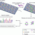
Randomization scheme. R randomization, ECC epirubicin, cisplatin, capecitabine (Adapted from Neo-adjuvant chemotherapy followed by surgery and chemotherapy or by surgery and chemoradiotherapy for patients with resectable gastric cancer (CRITICS). (2011), Dikken JL et al. [18])
In conclusion, postoperative adjuvant chemoradiotherapy should be selectively recommended for patients with resectable locally advanced gastric cancer.
7.2.2.3 Palliative Radiotherapy
Patients with late-stage gastric cancer always develop peritoneal carcinomatosis with symptoms of bleeding, pain, and intestinal obstruction, severely affecting the quality of life. Systemic chemotherapy is associated with low efficacy, higher incidence of toxicities, and poor tolerance. In contrast, local radiotherapy could alleviate symptoms and improve quality of life.
Chaw et al. evaluated the outcomes of 52 patients with gastric cancer bleeding who had been treated with palliative radiotherapy with hemostatic intent. Thirty-nine patients (75%) received single 8 Gy fraction, while 13 patients (25%) received 20 Gy in five daily fractions. The need for transfusion was evaluable in 44 patients, and the response rate was 50%, with fewer requirements for blood transfusions within 4 weeks of radiotherapy. The overall median survival (from treatment end to death) was 160 days (95% CI of 119–201 days), and the 1-year survival rate was 15% [19].
In a retrospective study, Tey et al. reviewed 115 patients that underwent palliative RT for index symptoms of gastric bleeding, obstruction, and pain. Dose fractionation regimens ranged from an 8 Gy single fraction to 40 Gy in 16 fractions. Response rates for bleeding, obstruction, and pain were 80.6% (83/103), 52.9% (9/17), and 45.5% (5/11), respectively. Median survival was significantly longer in patients who responded to RT compared with patients who did not (113.5 vs. 47 days, p < 0.001) [20].
7.3 The Technology of Radiotherapy for Gastric Cancer
Many abdominal organs such as the small intestine, liver, and kidneys are highly susceptible to radiotherapy-induced damage. Moreover, gastric cancer is associated with poor radiation sensitivity. Thus, it is necessary to take precautions to reduce radiation damage and to improve radiotherapy response.
7.3.1 The Range of Radiotherapy
According to the 2016 version of the NCCN Guidelines, the range of radiotherapy should be determined based on the location of tumor lesions both for neoadjuvant radiotherapy and postoperative adjuvant radiotherapy (Table 7.1).
Table 7.1
The range of radiotherapy for gastric cancer
Tumor site | Radiotherapy mode | CTV primary | CTV node |
|---|---|---|---|
Esophagogastric junction and upper 1/3 gastric | Neoadjuvant | Primary tumor and 3–5 cm range of proximal esophageal | Perigastric, peritoneal, splenic, hepatic portal lymph node area |
Adjuvant | Tumor bed and 3–5 cm range of proximal esophageal | Perigastric, peritoneal, splenic, hepatic portal lymph node area | |
Middle 1/3 gastric | Neoadjuvant | Primary tumor and peripheral subclinical lesions | Perigastric, pancreatic, peritoneal, splenic, hepatic portal, and pancreaticoduodenal lymph node area |
Adjuvant | Tumor bed (according to preoperative imaging scan or placement of the peptide clip), residual stomach would be in the CTV if the patient tolerate | Perigastric, pancreatic, peritoneal, splenic, hepatic portal, and pancreaticoduodenal lymph node area | |
Lower 1/3 gastric | Neoadjuvant | Primary tumor and peripheral subclinical lesions (if the tumor invasion of the stomach—duodenum—should include the first and second part of the duodenum) | Perigastric, pancreatic, peritoneal, hepatic portal, and pancreaticoduodenal lymph node area |
Adjuvant | Tumor bed and 3–5 cm range of duodenum | Perigastric, pancreatic, peritoneal, hepatic portal, and pancreaticoduodenal lymph node area |
Surgery may change the original anatomy, making it more difficult to sketch the postoperative lymphatic drainage area. Haijun et al. performed a phase II clinical study to determine CTV of adjuvant radiotherapy after radical surgery. The study demonstrated that the target area should include residual stomach, anastomosis, tumor bed, as well as the lymphatic drainage area. As lymph nodes are often associated with crucial blood vessels, the radiation target of the regional lymphatic drainage area can be determined based on remaining gastric and perigastric blood vessels. The most common grade 3–4 adverse event observed after adjuvant radiotherapy for gastric cancer was neutropenia (14.8%), while common grade 1–2 toxicities included neutropenia, nausea, and anemia. No treatment-related deaths occurred during the treatment period. The 3-year local recurrence-free survival rate was 91.1%, while the 3-year disease-free survival and overall survival rates were 70.2% and 81.6%, respectively. Eight patients developed peritoneal or distant metastasis [21].
7.3.2 The Dose of Radiotherapy
Due to the tolerable dose limit of abdominal organs such as the small intestine, liver, and kidneys, the maximum dose of neoadjuvant and postoperative adjuvant radiotherapy for gastric cancer is limited to approximately 50 Gy. The recommended dose is 1.8 Gy per fraction, with a total dose of 45–50.4 Gy. In case of positive margins or significant residual lesions after surgery, the radiation dose can be increased depending on the tolerable dose of the organ at risk near the target. Therefore, a radical effect of radiation therapy is difficult to achieve in patients with significant residual or intra-abdominal metastases.
7.3.3 The Tolerable Dose of Normal Tissue (Table 7.2)
Table 7.2
The tolerable dose of intra-abdominal organs
Organ | Tolerable dose |
|---|---|
Spinal cord | ≤40 Gy |
Liver | 60% of the liver volume ≤30 Gy |
Kidney | 33% of one side kidney volume ≤22.5 Gy, 33% of the other side kidney volume ≤45 Gy |
Small intestine | ≤54 Gy |
7.3.4 The Technique of Radiotherapy
7.3.4.1 Three-Dimensional Conformal Intensity-Modulated Radiotherapy
Radiation therapy has developed from two-dimensional to three-dimensional or even four-dimensional. In a retrospective study, Lee et al. investigated the dosimetric and clinical influence of three-dimensional (3D, CT-based) simulation versus conventional two-dimensional (2D)-based simulation in postoperative chemoradiotherapy in patients suffering from advanced gastric cancer. The 3D group showed better dose-volume histogram profiles compared to the 2D group for all dosimetric parameters, including the spinal cord, liver, duodenum, kidneys, and bowel [22].
Three-dimensional conformal radiotherapy (3DCRT) and intensity-modulated radiotherapy (IMRT) were performed using multi-field irradiation. The shape of each field was consistent with tumor shape. Additionally, the radiation dose was adjusted based on tumor cell density. For organs with a limited tolerable dose of radiation such as the spinal cord as well as abdominal organs including the liver, kidneys, and small intestine, the use of three-dimensional conformal intensity-modulated radiotherapy, the cornerstone of radiotherapy technology, is superior to two-dimensional radiotherapy, with better protection of normal abdominal tissue, a reduced frequency of adverse reactions, and improvement of the radiation dose accuracy. Tey’s study used three-dimensional conformal radiotherapy techniques to protect the normal tissue. The grade 3 adverse reaction rate was a mere 2.6%, and the therapy was well tolerated [20].
Intensity-modulated radiation therapy is based on reverse dose calculation. The target dose optimization is more reasonable, and the dose conformability is better resulting in improved protection of healthy tissue. In another study, Hawrylewicz et al. compared three-dimensional conformal radiotherapy with intensity-modulated radiotherapy in 25 patients with gastric cancer (adenocarcinoma T1–T4, N0–N3, and GI–GIII, according to AJCC). The area of clinical target volume (CTV) included a gastric tumor and 5 cm surrounding margins as well as the regional lymph nodes: perigastric, celiac trunk, pancreaticoduodenal, splenic, supra-pancreatic, portal vein, and para-aortic. Planning target volume (PTV) was determined by adding a 1 cm margin around the CTV. The planned total preoperative radiotherapy dose was 45 Gy administered in 25 fractions. During chemotherapy, 325 mg/m2 5-fluorouracil was applied (days 1–5). IMRT technology was used for treatment, and multiple CRT plans were made for comparison. The results of the study showed that the IMRT plan in the CTV conformity and homogeneity is better than the CRT plan (Table 7.3) [23].
Table 7.3
Comparison of conformity index and homogeneity index between IMRT and CRT
Two-field CRT | Three-field CRT | Four-field CRT | IMRT | |
|---|---|---|---|---|
Homogeneity index | 1.118 | 1.117 | 1.089 | 1.087 |
Conformity index | 1.115 | 1.118 | 1.088 | 1.082 |
7.3.4.2 Volumetric Intensity-Modulated Arc Therapy
Volumetric intensity-modulated arc therapy (VMAT) is one of the most advanced radiotherapy techniques. In VMAT, a computer controls the speed of the linear accelerator multi-leaf collimator velocity movement, the speed of frame rotation, and the dose rate. As a result, the tumor area is more accurately irradiated, while the surrounding healthy tissue only receives minimal radiation. This technique meets the goal of killing the tumor effectively and protecting the surrounding normal tissue.
VMAT technology incorporates various features: (1) a more even dose distribution within the target site through the use of rotating arc irradiation technology. This technology decreases the duration of treatment and improves treatment efficiency. (2) Rotating irradiation allows not only multi-leaf grating but also a dynamic dose rate change, making it easier to adjust the radiation field dose. (3) The current VMAT technology is equipped with image guidance function, making radiation therapy more accurate.
Currently, only two devices of this kind are in existence: the US Varian’s RapidArc and Sweden’s Elekta VMAT. Zhang et al. compared dose distributions of RapidArc (RA), static gantry intensity-modulated radiotherapy (IMRT), and three-dimensional conformal radiotherapy (3DCRT) as adjuvant radiotherapy modalities for the treatment of gastric cancer. The study included 15 patients with gastric cancer that underwent limited lymphadenectomy of perigastric lymph nodes. The CTV included the anastomosis, tumor bed, and regional lymph nodes. The PTV was defined as a uniform 5 mm expansion of the CTV. The liver, both kidneys, spinal cord, small intestine, heart, and other OAR were delineated. Dosimetric values for a total dose of 45 Gy/25f were calculated for each of the three modalities: the RapidArc, IMRT, and 3DCRT. The results demonstrated the following:
- (1)
PTV dose uniformity: IMRT and RapidArc were superior to 3DCRT, with RapidArc exhibiting the best dose uniformity.
- (2)
Dose of OAR: RapidArc excelled at liver and kidney protection compared to IMRT or 3DCRT.
- (3)
RapidArc significantly reduced the accelerator output dose, with a reduction of 42.5% compared with IMRT.
Furthermore, the dose rate of RapidArc can total 600 MU/min, which significantly decreases treatment time to half the time required for IMRT treatment. In conclusion, RapidArc has a significant advantage over both IMRT and 3DCRT [24].
7.3.4.3 Helical Tomotherapy
Helical tomotherapy (TOMO), the latest generation of radiation therapy equipment, was developed by the University of Wisconsin-Madison and TomoTherapy company. Known for its more precise, larger scope, and more complex treatment, it is a perfect fusion of CT and linear accelerator. TOMO uses the same X-ray source for both treatment and image guidance, thus avoiding the mechanical error of two sources of X-ray and improving the accuracy of the image guidance function. Like a CT scan, TOMO can complete a 160 cm range of irradiation in one session, without requiring multiple isocenter and repetition. TOMO uses the rotating irradiation approach to ensure even dose distribution. The first helical tomotherapy machine received FDA certification in 2002. Today, TOMO has become the mainstream equipment for radiotherapy.
In one study, Dahele et al. compared different types of radiotherapy in postoperative adjuvant radiotherapy in gastric cancer patients. Results showed that TOMO treatment was superior to 2F-CRT, 5F-CRT, and IMRT in the homogeneity of PTV. For the protection of normal tissues such as the kidneys and liver, TOMO was shown to perform comparably to IMRT and much better than CRT for its really lower V20 or V30, giving in an obvious advantage over traditional radiotherapy technology.
TOMO treatment was shown to be superior to CRT or IMRT not only in cases of extensive peritoneal metastasis, a common occurrence in patients with gastric cancer, but also in cases of ovarian cancer (Table 7.4) [25–27]. In this treatment regimen, the whole abdominal area was irradiated using TOMO. CTV included the whole peritoneal cavity, extending from the diaphragm to the Douglas cavity, and the pelvic and para-aortic node regions. These studies indicate an alternative treatment for patients with extensive peritoneal metastasis.
Table 7.4
Studies for whole abdominal irradiation using TOMO
Author | Year | Number of cases | Treatment purposes | Irradiation method | Radiation dose | Results | Adverse events |
|---|---|---|---|---|---|---|---|
Rochet N | 2015 | 16 | Adjuvant radiotherapy | TOMO or step-and-shoot IMRT | 30 Gy/20f | Median RFS was 27.6 months, median OS was 42.1 months | Grade 3 toxicities were diarrhea (25%), leucopenia (19%), nausea/vomiting (6%), and thrombocytopenia (6%) |
Shetty UM | 2013 | 8 | Palliative radiotherapy
Stay updated, free articles. Join our Telegram channel
Full access? Get Clinical Tree
 Get Clinical Tree app for offline access

|


